1UPT

Structure of a complex of the golgin-245 GRIP domain with Arl1


Experimental Data Snapshot

  • Method: X-RAY DIFFRACTION
  • Resolution: 1.70 Å
  • R-Value Free: 0.252 
  • R-Value Work: 0.221 
  • R-Value Observed: 0.222 

wwPDB Validation   3D Report Full Report


Ligand Structure Quality Assessment 


This is version 1.4 of the entry. See complete history


Literature

Structural Basis for Arl1-Dependent Targeting of Homodimeric Grip Domains to the Golgi Apparatus

Panic, B.Perisic, O.Veprintsev, D.B.Williams, R.L.Munro, S.

(2003) Mol Cell 12: 863

  • DOI: https://doi.org/10.1016/s1097-2765(03)00356-3
  • Primary Citation of Related Structures:  
    1UPT

  • PubMed Abstract: 

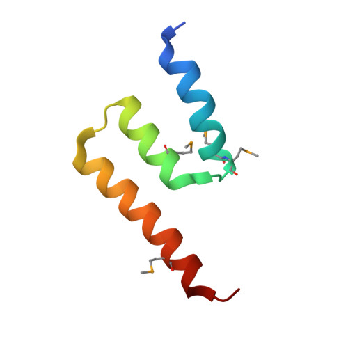
    Golgins are large coiled-coil proteins that play a role in Golgi structure and vesicle traffic. The Arf-like GTPase Arl1 regulates the translocation of GRIP domain-containing golgins to Golgi membranes. We report here the 1.7 A resolution structure of human Arl1-GTP in a complex with the GRIP domain of golgin-245. The structure reveals that the GRIP domain consists of an S-shaped arrangement of three helices. The domain forms a homodimer that binds two Arl1-GTPs using two helices from each monomer. The structure is consistent with golgin-245 forming parallel coiled-coils and suggests how Arl1-GTP/GRIP complexes interact with Golgi membranes via the N termini of Arl1-GTP and the C-terminal tails of the GRIP domains. In cells, bivalent association with Arl1-GTP would increase residence time of the golgins on Golgi membranes. Despite no conservation of sequence, topology, or even helical direction, several other effectors form similar interactions with small GTPases via a pair of alpha helices, suggesting a common structural basis for effector recognition.


  • Organizational Affiliation

    MRC Laboratory of Molecular Biology, MRC Centre, Hills Road, Cambridge CB2 2QH, United Kingdom.


Macromolecules
Find similar proteins by:  (by identity cutoff)  |  3D Structure
Entity ID: 1
MoleculeChains Sequence LengthOrganismDetailsImage
ADP-RIBOSYLATION FACTOR-LIKE PROTEIN 1
A, C, E, G
171Homo sapiensMutation(s): 0 
UniProt & NIH Common Fund Data Resources
Find proteins for P40616 (Homo sapiens)
Explore P40616 
Go to UniProtKB:  P40616
PHAROS:  P40616
GTEx:  ENSG00000120805 
Entity Groups  
Sequence Clusters30% Identity50% Identity70% Identity90% Identity95% Identity100% Identity
UniProt GroupP40616
Sequence Annotations
Expand
  • Reference Sequence
Find similar proteins by:  (by identity cutoff)  |  3D Structure
Entity ID: 2
MoleculeChains Sequence LengthOrganismDetailsImage
GOLGI AUTOANTIGEN, GOLGIN SUBFAMILY A MEMBER 4
B, D, F, H
60Homo sapiensMutation(s): 7 
UniProt & NIH Common Fund Data Resources
Find proteins for Q13439 (Homo sapiens)
Explore Q13439 
Go to UniProtKB:  Q13439
PHAROS:  Q13439
GTEx:  ENSG00000144674 
Entity Groups  
Sequence Clusters30% Identity50% Identity70% Identity90% Identity95% Identity100% Identity
UniProt GroupQ13439
Sequence Annotations
Expand
  • Reference Sequence
Experimental Data & Validation

Experimental Data

  • Method: X-RAY DIFFRACTION
  • Resolution: 1.70 Å
  • R-Value Free: 0.252 
  • R-Value Work: 0.221 
  • R-Value Observed: 0.222 
  • Space Group: P 1 21 1
Unit Cell:
Length ( Å )Angle ( ˚ )
a = 72.497α = 90
b = 89.711β = 110.69
c = 72.534γ = 90
Software Package:
Software NamePurpose
MOSFLMdata reduction
SCALAdata scaling
SnBphasing
SHARPphasing
REFMACrefinement

Structure Validation

View Full Validation Report


Ligand Structure Quality Assessment 


Entry History 

Deposition Data

Revision History  (Full details and data files)

  • Version 1.0: 2003-10-30
    Type: Initial release
  • Version 1.1: 2011-05-08
    Changes: Version format compliance
  • Version 1.2: 2011-07-13
    Changes: Version format compliance
  • Version 1.3: 2017-07-05
    Changes: Refinement description
  • Version 1.4: 2019-10-23
    Changes: Data collection, Database references, Derived calculations, Other