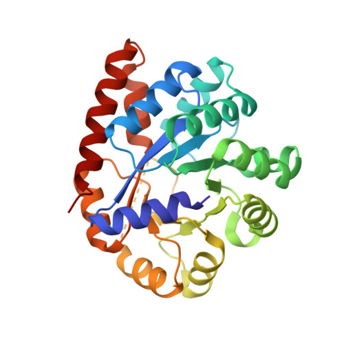
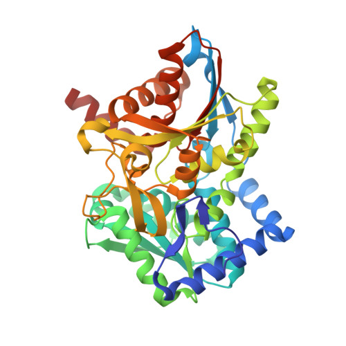
1K7E

CRYSTAL STRUCTURE OF WILD-TYPE TRYPTOPHAN SYNTHASE COMPLEXED WITH N-[1H-INDOL-3-YL-ACETYL]GLYCINE ACID


Experimental Data Snapshot

  • Method: X-RAY DIFFRACTION
  • Resolution: 2.30 Å
  • R-Value Free: 0.244 
  • R-Value Work: 0.167 
  • R-Value Observed: 0.171 

Starting Model: experimental
View more details

wwPDB Validation   3D Report Full Report


Ligand Structure Quality Assessment 


This is version 1.3 of the entry. See complete history


Literature

Crystal structures of a new class of allosteric effectors complexed to tryptophan synthase.

Weyand, M.Schlichting, I.Marabotti, A.Mozzarelli, A.

(2002) J Biol Chem 277: 10647-10652

  • DOI: https://doi.org/10.1074/jbc.M111285200
  • Primary Citation of Related Structures:  
    1K3U, 1K7E, 1K7F

  • PubMed Abstract: 

    Tryptophan synthase is a bifunctional alpha(2)beta(2) complex catalyzing the last two steps of l-tryptophan biosynthesis. The natural substrates of the alpha-subunit indole- 3-glycerolphosphate and glyceraldehyde-3-phosphate, and the substrate analogs indole-3-propanolphosphate and dl-alpha-glycerol-3-phosphate are allosteric effectors of the beta-subunit activity. It has been shown recently, that the indole-3-acetyl amino acids indole-3-acetylglycine and indole-3-acetyl-l-aspartic acid are both alpha-subunit inhibitors and beta-subunit allosteric effectors, whereas indole-3-acetyl-l-valine is only an alpha-subunit inhibitor (Marabotti, A., Cozzini, P., and Mozzarelli, A. (2000) Biochim. Biophys. Acta 1476, 287-299). The crystal structures of tryptophan synthase complexed with indole-3-acetylglycine and indole-3-acetyl-l-aspartic acid show that both ligands bind to the active site such that the carboxylate moiety is positioned similarly as the phosphate group of the natural substrates. As a consequence, the residues of the alpha-active site that interact with the ligands are the same as observed in the indole 3-glycerolphosphate-enzyme complex. Ligand binding leads to closure of loop alphaL6 of the alpha-subunit, a key structural element of intersubunit communication. This is in keeping with the allosteric role played by these compounds. The structure of the enzyme complex with indole-3-acetyl-l-valine is quite different. Due to the hydrophobic lateral chain, this molecule adopts a new orientation in the alpha-active site. In this case, closure of loop alphaL6 is no longer observed, in agreement with its functioning only as an inhibitor of the alpha-subunit reaction.


  • Organizational Affiliation

    Max-Planck-Institut für Molekulare Physiologie, Abteilung für Physikalische Biochemie, D-44227 Dortmund, Germany.


Macromolecules
Find similar proteins by:  (by identity cutoff)  |  3D Structure
Entity ID: 1
MoleculeChains Sequence LengthOrganismDetailsImage
TRYPTOPHAN SYNTHASE ALPHA CHAIN268Salmonella enterica subsp. enterica serovar TyphimuriumMutation(s): 0 
Gene Names: TRPA/TRPB
EC: 4.2.1.20
UniProt
Find proteins for P00929 (Salmonella typhimurium (strain LT2 / SGSC1412 / ATCC 700720))
Explore P00929 
Go to UniProtKB:  P00929
Entity Groups  
Sequence Clusters30% Identity50% Identity70% Identity90% Identity95% Identity100% Identity
UniProt GroupP00929
Sequence Annotations
Expand
  • Reference Sequence
Find similar proteins by:  (by identity cutoff)  |  3D Structure
Entity ID: 2
MoleculeChains Sequence LengthOrganismDetailsImage
TRYPTOPHAN SYNTHASE BETA CHAIN396Salmonella enterica subsp. enterica serovar TyphimuriumMutation(s): 0 
Gene Names: TRPA/TRPB
EC: 4.2.1.20
UniProt
Find proteins for P0A2K1 (Salmonella typhimurium (strain LT2 / SGSC1412 / ATCC 700720))
Explore P0A2K1 
Go to UniProtKB:  P0A2K1
Entity Groups  
Sequence Clusters30% Identity50% Identity70% Identity90% Identity95% Identity100% Identity
UniProt GroupP0A2K1
Sequence Annotations
Expand
  • Reference Sequence
Experimental Data & Validation

Experimental Data

  • Method: X-RAY DIFFRACTION
  • Resolution: 2.30 Å
  • R-Value Free: 0.244 
  • R-Value Work: 0.167 
  • R-Value Observed: 0.171 
  • Space Group: C 1 2 1
Unit Cell:
Length ( Å )Angle ( ˚ )
a = 182.651α = 90
b = 59.081β = 94.55
c = 67.3γ = 90
Software Package:
Software NamePurpose
CNSrefinement
REFMACrefinement
XDSdata reduction
XSCALEdata scaling
CNSphasing

Structure Validation

View Full Validation Report


Ligand Structure Quality Assessment 


Entry History 

Deposition Data

Revision History  (Full details and data files)

  • Version 1.0: 2002-07-10
    Type: Initial release
  • Version 1.1: 2008-04-27
    Changes: Version format compliance
  • Version 1.2: 2011-07-13
    Changes: Non-polymer description, Version format compliance
  • Version 1.3: 2023-08-16
    Changes: Data collection, Database references, Derived calculations, Refinement description