1DGH

HUMAN ERYTHROCYTE CATALASE 3-AMINO-1,2,4-TRIAZOLE COMPLEX


Experimental Data Snapshot

  • Method: X-RAY DIFFRACTION
  • Resolution: 2.00 Å
  • R-Value Free: 0.210 
  • R-Value Work: 0.173 
  • R-Value Observed: 0.177 

wwPDB Validation   3D Report Full Report


Ligand Structure Quality Assessment 


This is version 1.2 of the entry. See complete history


Literature

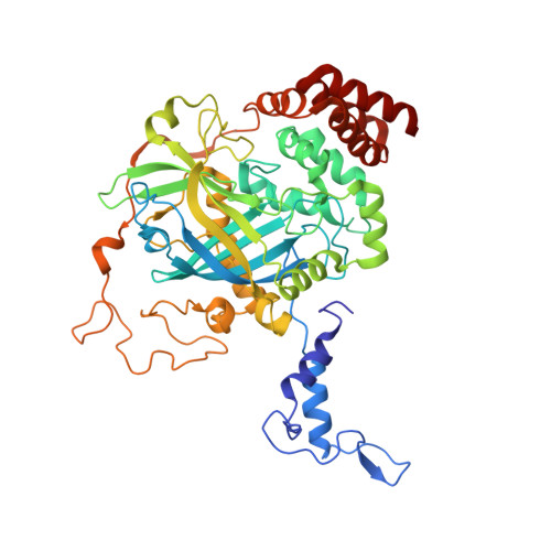
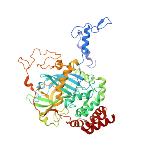
Active and inhibited human catalase structures: ligand and NADPH binding and catalytic mechanism.

Putnam, C.D.Arvai, A.S.Bourne, Y.Tainer, J.A.

(2000) J Mol Biol 296: 295-309

  • DOI: 10.1006/jmbi.1999.3458
  • Primary Citation of Related Structures:  
    1DGB, 1DGF, 1DGG, 1DGH

  • PubMed Abstract: 
  • Human catalase is an heme-containing peroxisomal enzyme that breaks down hydrogen peroxide to water and oxygen; it is implicated in ethanol metabolism, inflammation, apoptosis, aging and cancer. The 1. 5 A resolution human enzyme structure, both with and without bound NADPH, establishes the conserved features of mammalian catalase fold and assembly, implicates Tyr370 as the tyrosine radical, suggests the structural basis for redox-sensitive binding of cognate mRNA via the catalase NADPH binding site, and identifies an unexpectedly substantial number of water-mediated domain contacts ...

    Human catalase is an heme-containing peroxisomal enzyme that breaks down hydrogen peroxide to water and oxygen; it is implicated in ethanol metabolism, inflammation, apoptosis, aging and cancer. The 1. 5 A resolution human enzyme structure, both with and without bound NADPH, establishes the conserved features of mammalian catalase fold and assembly, implicates Tyr370 as the tyrosine radical, suggests the structural basis for redox-sensitive binding of cognate mRNA via the catalase NADPH binding site, and identifies an unexpectedly substantial number of water-mediated domain contacts. A molecular ruler mechanism based on observed water positions in the 25 A-long channel resolves problems for selecting hydrogen peroxide. Control of water-mediated hydrogen bonds by this ruler selects for the longer hydrogen peroxide and explains the paradoxical effects of mutations that increase active site access but lower catalytic rate. The heme active site is tuned without compromising peroxide binding through a Tyr-Arg-His-Asp charge relay, arginine residue to heme carboxylate group hydrogen bonding, and aromatic stacking. Structures of the non-specific cyanide and specific 3-amino-1,2, 4-triazole inhibitor complexes of human catalase identify their modes of inhibition and help reveal the catalytic mechanism of catalase. Taken together, these resting state and inhibited human catalase structures support specific, structure-based mechanisms for the catalase substrate recognition, reaction and inhibition and provide a molecular basis for understanding ethanol intoxication and the likely effects of human polymorphisms.


    Organizational Affiliation

    Department of Molecular Biology, Skaggs Institute for Chemical Biology, The Scripps Research Institute, MB 4, 10550 North Torrey Pines Road, La Jolla, CA 92037, USA.


Macromolecules
Find similar proteins by:  (by identity cutoff)  |  3D Structure
Entity ID: 1
MoleculeChainsSequence LengthOrganismDetailsImage
PROTEIN (CATALASE)A, C498Homo sapiensMutation(s): 0 
Gene Names: CAT
EC: 1.11.1.6
UniProt & NIH Common Fund Data Resources
Find proteins for P04040 (Homo sapiens)
Explore P04040 
Go to UniProtKB:  P04040
PHAROS:  P04040
Entity Groups  
Sequence Clusters30% Identity50% Identity70% Identity90% Identity95% Identity100% Identity
UniProt GroupP04040
Protein Feature View
Expand
  • Reference Sequence
Find similar proteins by:  (by identity cutoff)  |  3D Structure
Entity ID: 2
MoleculeChainsSequence LengthOrganismDetailsImage
PROTEIN (CATALASE)B, D498Homo sapiensMutation(s): 1 
Gene Names: CAT
EC: 1.11.1.6
UniProt & NIH Common Fund Data Resources
Find proteins for P04040 (Homo sapiens)
Explore P04040 
Go to UniProtKB:  P04040
PHAROS:  P04040
Entity Groups  
Sequence Clusters30% Identity50% Identity70% Identity90% Identity95% Identity100% Identity
UniProt GroupP04040
Protein Feature View
Expand
  • Reference Sequence
Small Molecules
Ligands 2 Unique
IDChainsName / Formula / InChI Key2D Diagram3D Interactions
NDP
Query on NDP

Download Ideal Coordinates CCD File 
F [auth A],
I [auth C]
NADPH DIHYDRO-NICOTINAMIDE-ADENINE-DINUCLEOTIDE PHOSPHATE
C21 H30 N7 O17 P3
ACFIXJIJDZMPPO-NNYOXOHSSA-N
 Ligand Interaction
HEM
Query on HEM

Download Ideal Coordinates CCD File 
E [auth A],
G [auth B],
H [auth C],
J [auth D]
PROTOPORPHYRIN IX CONTAINING FE
C34 H32 Fe N4 O4
KABFMIBPWCXCRK-RGGAHWMASA-L
 Ligand Interaction
Modified Residues  1 Unique
IDChainsTypeFormula2D DiagramParent
3AH
Query on 3AH
B, D L-PEPTIDE LINKINGC8 H11 N7 O2HIS
Experimental Data & Validation

Experimental Data

  • Method: X-RAY DIFFRACTION
  • Resolution: 2.00 Å
  • R-Value Free: 0.210 
  • R-Value Work: 0.173 
  • R-Value Observed: 0.177 
  • Space Group: P 21 21 21
Unit Cell:
Length ( Å )Angle ( ˚ )
a = 84.028α = 90
b = 140.639β = 90
c = 231.346γ = 90
Software Package:
Software NamePurpose
AMoREphasing
X-PLORrefinement
DENZOdata reduction
SCALEPACKdata scaling

Structure Validation

View Full Validation Report


Ligand Structure Quality Assessment 


Entry History 

Deposition Data

Revision History  (Full details and data files)

  • Version 1.0: 2000-02-17
    Type: Initial release
  • Version 1.1: 2008-04-27
    Changes: Version format compliance
  • Version 1.2: 2011-07-13
    Changes: Version format compliance