1ZUN

Crystal Structure of a GTP-Regulated ATP Sulfurylase Heterodimer from Pseudomonas syringae


Experimental Data Snapshot

  • Method: X-RAY DIFFRACTION
  • Resolution: 2.70 Å
  • R-Value Free: 0.276 
  • R-Value Work: 0.222 
  • R-Value Observed: 0.224 

wwPDB Validation   3D Report Full Report


Ligand Structure Quality Assessment 


This is version 1.2 of the entry. See complete history


Literature

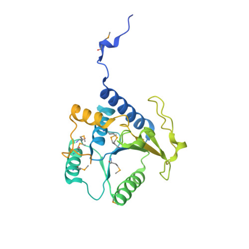
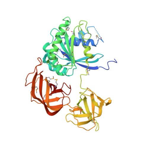
Molecular basis for g protein control of the prokaryotic ATP sulfurylase.

Mougous, J.D.Lee, D.H.Hubbard, S.C.Schelle, M.W.Vocadlo, D.J.Berger, J.M.Bertozzi, C.R.

(2006) Mol Cell 21: 109-122

  • DOI: https://doi.org/10.1016/j.molcel.2005.10.034
  • Primary Citation of Related Structures:  
    1ZUN

  • PubMed Abstract: 
  • Sulfate assimilation is a critical component of both primary and secondary metabolism. An essential step in this pathway is the activation of sulfate through adenylation by the enzyme ATP sulfurylase (ATPS), forming adenosine 5'-phosphosulfate (APS). Proteobacterial ATPS overcomes this energetically unfavorable reaction by associating with a regulatory G protein, coupling the energy of GTP hydrolysis to APS formation ...

    Sulfate assimilation is a critical component of both primary and secondary metabolism. An essential step in this pathway is the activation of sulfate through adenylation by the enzyme ATP sulfurylase (ATPS), forming adenosine 5'-phosphosulfate (APS). Proteobacterial ATPS overcomes this energetically unfavorable reaction by associating with a regulatory G protein, coupling the energy of GTP hydrolysis to APS formation. To discover the molecular basis of this unusual role for a G protein, we biochemically characterized and solved the X-ray crystal structure of a complex between Pseudomonas syringae ATPS (CysD) and its associated regulatory G protein (CysN). The structure of CysN*D shows the two proteins in tight association; however, the nucleotides bound to each subunit are spatially segregated. We provide evidence that conserved switch motifs in the G domain of CysN allosterically mediate interactions between the nucleotide binding sites. This structure suggests a molecular mechanism by which conserved G domain architecture is used to energetically link GTP turnover to the production of an essential metabolite.


    Organizational Affiliation

    Howard Hughes Medical Institute, University of California, Berkeley, Berkeley, California 94720, USA.


Macromolecules
Find similar proteins by:  (by identity cutoff)  |  3D Structure
Entity ID: 1
MoleculeChainsSequence LengthOrganismDetailsImage
Sulfate adenylyltransferase subunit 2A325Pseudomonas syringaeMutation(s): 8 
Gene Names: cysD
EC: 2.7.7.4
UniProt
Find proteins for Q87WW0 (Pseudomonas syringae pv. tomato (strain ATCC BAA-871 / DC3000))
Explore Q87WW0 
Go to UniProtKB:  Q87WW0
Entity Groups  
Sequence Clusters30% Identity50% Identity70% Identity90% Identity95% Identity100% Identity
UniProt GroupQ87WW0
Protein Feature View
Expand
  • Reference Sequence
Find similar proteins by:  (by identity cutoff)  |  3D Structure
Entity ID: 2
MoleculeChainsSequence LengthOrganismDetailsImage
sulfate adenylate transferase, subunit 1/adenylylsulfate kinaseB434Pseudomonas syringae pv. tomato str. DC3000Mutation(s): 12 
Gene Names: cysNCcysNCPSPTO_4432
EC: 2.7.7.4
UniProt
Find proteins for Q87WW1 (Pseudomonas syringae pv. tomato (strain ATCC BAA-871 / DC3000))
Explore Q87WW1 
Go to UniProtKB:  Q87WW1
Entity Groups  
Sequence Clusters30% Identity50% Identity70% Identity90% Identity95% Identity100% Identity
UniProt GroupQ87WW1
Protein Feature View
Expand
  • Reference Sequence
Small Molecules
Ligands 4 Unique
IDChainsName / Formula / InChI Key2D Diagram3D Interactions
AGS
Query on AGS

Download Ideal Coordinates CCD File 
E [auth A]PHOSPHOTHIOPHOSPHORIC ACID-ADENYLATE ESTER
C10 H16 N5 O12 P3 S
NLTUCYMLOPLUHL-KQYNXXCUSA-N
 Ligand Interaction
GDP
Query on GDP

Download Ideal Coordinates CCD File 
F [auth B]GUANOSINE-5'-DIPHOSPHATE
C10 H15 N5 O11 P2
QGWNDRXFNXRZMB-UUOKFMHZSA-N
 Ligand Interaction
MG
Query on MG

Download Ideal Coordinates CCD File 
C [auth A]MAGNESIUM ION
Mg
JLVVSXFLKOJNIY-UHFFFAOYSA-N
 Ligand Interaction
NA
Query on NA

Download Ideal Coordinates CCD File 
D [auth A]SODIUM ION
Na
FKNQFGJONOIPTF-UHFFFAOYSA-N
 Ligand Interaction
Modified Residues  1 Unique
IDChainsTypeFormula2D DiagramParent
MSE
Query on MSE
A L-PEPTIDE LINKINGC5 H11 N O2 SeMET
Experimental Data & Validation

Experimental Data

  • Method: X-RAY DIFFRACTION
  • Resolution: 2.70 Å
  • R-Value Free: 0.276 
  • R-Value Work: 0.222 
  • R-Value Observed: 0.224 
  • Space Group: H 3
Unit Cell:
Length ( Å )Angle ( ˚ )
a = 110.072α = 90
b = 110.072β = 90
c = 171.002γ = 120
Software Package:
Software NamePurpose
REFMACrefinement
HKL-2000data reduction
SCALEPACKdata scaling
SOLVEphasing

Structure Validation

View Full Validation Report


Ligand Structure Quality Assessment 


Entry History 

Deposition Data

Revision History  (Full details and data files)

  • Version 1.0: 2006-01-17
    Type: Initial release
  • Version 1.1: 2008-04-30
    Changes: Version format compliance
  • Version 1.2: 2011-07-13
    Changes: Version format compliance