1ZE8 | pdb_00001ze8

Carbonic anhydrase II in complex with a membrane-impermeant sulfonamide inhibitor


Experimental Data Snapshot

  • Method: X-RAY DIFFRACTION
  • Resolution: 2.00 Å
  • R-Value Free: 
    0.251 (Depositor), 0.237 (DCC) 
  • R-Value Work: 
    0.204 (Depositor), 0.200 (DCC) 
  • R-Value Observed: 
    0.221 (Depositor) 

wwPDB Validation 3D Report Full Report

Validation slider image for 1ZE8

Ligand Structure Quality Assessment 


This is version 1.3 of the entry. See complete history

Literature

Carbonic anhydrase inhibitors: stacking with Phe131 determines active site binding region of inhibitors as exemplified by the X-ray crystal structure of a membrane-impermeant antitumor sulfonamide complexed with isozyme II

Menchise, V.De Simone, G.Alterio, V.Di Fiore, A.Pedone, C.Scozzafava, A.Supuran, C.T.

(2005) J Med Chem 48: 5721-5727

  • DOI: https://doi.org/10.1021/jm050333c
  • Primary Citation Related Structures: 
    1ZE8

  • PubMed Abstract: 

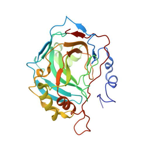
    Structure for the adduct of carbonic anhydrase II with 1-N-(4-sulfamoylphenyl-ethyl)-2,4,6-trimethylpyridinium perchlorate, a membrane-impermeant antitumor sulfonamide, is reported. The phenylethyl moiety fills the active site, making van der Waals interactions with side chains of Gln192, Val121, Phe131, Leu198, Thr200. The 2,4,6-trimethylpyridinium functionality is at van der Waals distance from the aliphatic chain of Ile91 being involved in strong offset face-to-face stacking with Phe131. Analyzing X-ray crystal structures of such adducts, two binding modes were observed: some inhibitors bind with their tail within the hydrophobic half of the active site, defined by residues Phe131, Val135, Leu198, Pro202, Leu204. Other derivatives bind with their tail in a different region, pointing toward the hydrophilic half and making strong parallel stacking with Phe131. This interaction orients the inhibitor toward the hydrophilic part of the active site. Impossibility to participate in it leads to its binding within the hydrophobic half. Such findings are relevant for designing better inhibitors targeting isozymes II, IX, and XII, some of which are overexpressed in hypoxic tumors.


  • Organizational Affiliation
    • Istituto di Biostrutture e Bioimmagini-CNR, via Mezzocannone 16, 80134 Naples, Italy.

Macromolecule Content 

  • Total Structure Weight: 29.96 kDa 
  • Atom Count: 2,295 
  • Modeled Residue Count: 258 
  • Deposited Residue Count: 259 
  • Unique protein chains: 1

Macromolecules

Find similar proteins by:|  3D Structure
Entity ID: 1
MoleculeChains  Sequence LengthOrganismDetailsImage
Carbonic anhydrase II259Homo sapiensMutation(s): 0 
EC: 4.2.1.1 (PDB Primary Data), 4.2.1.69 (UniProt)
UniProt & NIH Common Fund Data Resources
Find proteins for P00918 (Homo sapiens)
Explore P00918 
Go to UniProtKB:  P00918
PHAROS:  P00918
GTEx:  ENSG00000104267 
Entity Groups
Sequence Clusters30% Identity50% Identity70% Identity90% Identity95% Identity100% Identity
UniProt GroupP00918
Sequence Annotations
Expand
Reference Sequence

Experimental Data & Validation

Experimental Data

  • Method: X-RAY DIFFRACTION
  • Resolution: 2.00 Å
  • R-Value Free:  0.251 (Depositor), 0.237 (DCC) 
  • R-Value Work:  0.204 (Depositor), 0.200 (DCC) 
  • R-Value Observed: 0.221 (Depositor) 
Space Group: P 1 21 1
Unit Cell:
Length ( Å )Angle ( ˚ )
a = 42.17α = 90
b = 41.39β = 104.29
c = 72.04γ = 90
Software Package:
Software NamePurpose
DENZOdata reduction
SCALEPACKdata scaling
CNSrefinement
CNSphasing

Structure Validation

View Full Validation Report


Ligand Structure Quality Assessment 


Entry History 

Deposition Data

Revision History  (Full details and data files)

  • Version 1.0: 2005-10-18
    Type: Initial release
  • Version 1.1: 2008-04-30
    Changes: Version format compliance
  • Version 1.2: 2011-07-13
    Changes: Non-polymer description, Version format compliance
  • Version 1.3: 2024-03-13
    Changes: Data collection, Database references, Derived calculations