1YCD | pdb_00001ycd

Crystal structure of yeast FSH1/YHR049W, a member of the serine hydrolase family


Experimental Data Snapshot

  • Method: X-RAY DIFFRACTION
  • Resolution: 1.70 Å
  • R-Value Free: 
    0.202 (Depositor), 0.165 (DCC) 
  • R-Value Work: 
    0.166 (Depositor), 0.167 (DCC) 
  • R-Value Observed: 
    0.167 (Depositor) 

wwPDB Validation 3D Report Full Report

Validation slider image for 1YCD

Ligand Structure Quality Assessment 


This is version 1.4 of the entry. See complete history

Literature

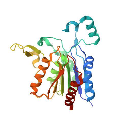
Crystal structure of yeast YHR049W/FSH1, a member of the serine hydrolase family.

Quevillon-Cheruel, S.Leulliot, N.Graille, M.Hervouet, N.Coste, F.Zelwer, C.Janin, J.Van Tilbeurgh, H.

(2005) Protein Sci 14: 1350-1356

  • DOI: https://doi.org/10.1110/ps.051415905
  • Primary Citation Related Structures: 
    1YCD

  • PubMed Abstract: 

    Yhr049w/FSH1 was recently identified in a combined computational and experimental proteomics analysis for the detection of active serine hydrolases in yeast. This analysis suggested that FSH1 might be a serine-type hydrolase belonging to the broad functional alphabeta-hydrolase superfamily. In order to get insight into the molecular function of this gene, it was targeted in our yeast structural genomics project. The crystal structure of the protein confirms that it contains a Ser/His/Asp catalytic triad that is part of a minimal alpha/beta-hydrolase fold. The architecture of the putative active site and analogies with other protein structures suggest that FSH1 may be an esterase. This finding was further strengthened by the unexpected presence of a compound covalently bound to the catalytic serine in the active site. Apparently, the enzyme was trapped with a reactive compound during the purification process.


  • Organizational Affiliation
    • Institut de Biochimie et de Biophysique Moléculaire et Cellulaire (CNRS-UMR 8619), Université Paris-Sud, Bâtiment 430, 91405 Orsay, France.

Macromolecule Content 

  • Total Structure Weight: 55.13 kDa 
  • Atom Count: 4,732 
  • Modeled Residue Count: 475 
  • Deposited Residue Count: 486 
  • Unique protein chains: 1

Macromolecules

Find similar proteins by:|  3D Structure
Entity ID: 1
MoleculeChains  Sequence LengthOrganismDetailsImage
Hypothetical 27.3 kDa protein in AAP1-SMF2 intergenic region
A, B
243Saccharomyces cerevisiaeMutation(s): 1 
Gene Names: YHR049w
EC: 3.1
UniProt
Find proteins for P38777 (Saccharomyces cerevisiae (strain ATCC 204508 / S288c))
Explore P38777 
Go to UniProtKB:  P38777
Entity Groups
Sequence Clusters30% Identity50% Identity70% Identity90% Identity95% Identity100% Identity
UniProt GroupP38777
Sequence Annotations
Expand
Reference Sequence

Experimental Data & Validation

Experimental Data

  • Method: X-RAY DIFFRACTION
  • Resolution: 1.70 Å
  • R-Value Free:  0.202 (Depositor), 0.165 (DCC) 
  • R-Value Work:  0.166 (Depositor), 0.167 (DCC) 
  • R-Value Observed: 0.167 (Depositor) 
Space Group: P 1
Unit Cell:
Length ( Å )Angle ( ˚ )
a = 46.133α = 102.85
b = 53.329β = 90.04
c = 64.271γ = 112.47
Software Package:
Software NamePurpose
REFMACrefinement
DENZOdata reduction
SCALEPACKdata scaling
SHARPphasing

Structure Validation

View Full Validation Report


Ligand Structure Quality Assessment 


Entry History 

Revision History  (Full details and data files)

  • Version 1.0: 2005-05-10
    Type: Initial release
  • Version 1.1: 2008-04-30
    Changes: Version format compliance
  • Version 1.2: 2011-07-13
    Changes: Version format compliance
  • Version 1.3: 2021-10-20
    Changes: Database references, Derived calculations
  • Version 1.4: 2024-10-30
    Changes: Data collection, Structure summary