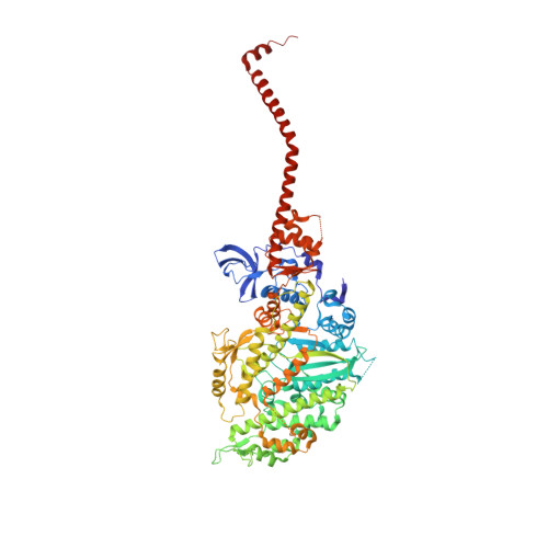
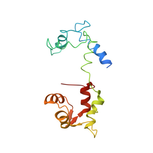
1SR6

Structure of nucleotide-free scallop myosin S1


Experimental Data Snapshot

  • Method: X-RAY DIFFRACTION
  • Resolution: 2.75 Å
  • R-Value Free: 0.286 
  • R-Value Work: 0.242 
  • R-Value Observed: 0.242 

wwPDB Validation   3D Report Full Report


This is version 1.2 of the entry. See complete history


Literature

Myosin subfragment 1 structures reveal a partially bound nucleotide and a complex salt bridge that helps couple nucleotide and actin binding.

Risal, D.Gourinath, S.Himmel, D.M.Szent-Gyorgyi, A.G.Cohen, C.

(2004) Proc Natl Acad Sci U S A 101: 8930-8935

  • DOI: 10.1073/pnas.0403002101
  • Primary Citation of Related Structures:  
    1S5G, 1SR6

  • PubMed Abstract: 
  • Structural studies of myosin have indicated some of the conformational changes that occur in this protein during the contractile cycle, and we have now observed a conformational change in a bound nucleotide as well. The 3.1-A x-ray structure of the scallop myosin head domain (subfragment 1) in the ADP-bound near-rigor state (lever arm =45 degrees to the helical actin axis) shows the diphosphate moiety positioned on the surface of the nucleotide-binding pocket, rather than deep within it as had been observed previously ...

    Structural studies of myosin have indicated some of the conformational changes that occur in this protein during the contractile cycle, and we have now observed a conformational change in a bound nucleotide as well. The 3.1-A x-ray structure of the scallop myosin head domain (subfragment 1) in the ADP-bound near-rigor state (lever arm =45 degrees to the helical actin axis) shows the diphosphate moiety positioned on the surface of the nucleotide-binding pocket, rather than deep within it as had been observed previously. This conformation strongly suggests a specific mode of entry and exit of the nucleotide from the nucleotide-binding pocket through the so-called "front door." In addition, using a variety of scallop structures, including a relatively high-resolution 2.75-A nucleotide-free near-rigor structure, we have identified a conserved complex salt bridge connecting the 50-kDa upper and N-terminal subdomains. This salt bridge is present only in crystal structures of muscle myosin isoforms that exhibit a strong reciprocal relationship (also known as coupling) between actin and nucleotide affinity.


    Related Citations: 
    • Crystal structure of scallop Myosin s1 in the pre-power stroke state to 2.6 a resolution: flexibility and function in the head
      Gourinath, S., Himmel, D.M., Brown, J.H., Reshetnikova, L., Szent-Gyorgyi, A.G., Cohen, C.
      (2003) Structure 11: 1621
    • Crystallographic findings on the internally uncoupled and near-rigor states of myosin: further insights into the mechanics of the motor
      Himmel, D.M., Gourinath, S., Reshetnikova, L., Shen, Y., Szent-Gyorgyi, A.G., Cohen, C.
      (2002) Proc Natl Acad Sci U S A 99: 12645

    Organizational Affiliation

    Rosenstiel Basic Medical Sciences Research Center, MS 029, Waltham, MA 02454-9110, USA.


Macromolecules
Find similar proteins by:  (by identity cutoff)  |  3D Structure
Entity ID: 1
MoleculeChainsSequence LengthOrganismDetailsImage
Myosin heavy chain, striated muscleA840Argopecten irradiansMutation(s): 0 
UniProt
Find proteins for P24733 (Argopecten irradians)
Explore P24733 
Go to UniProtKB:  P24733
Entity Groups  
Sequence Clusters30% Identity50% Identity70% Identity90% Identity95% Identity100% Identity
UniProt GroupP24733
Protein Feature View
Expand
  • Reference Sequence
Find similar proteins by:  (by identity cutoff)  |  3D Structure
Entity ID: 2
MoleculeChainsSequence LengthOrganismDetailsImage
Myosin regulatory light chain, striated adductor muscleB156Argopecten irradiansMutation(s): 0 
UniProt
Find proteins for P13543 (Argopecten irradians)
Explore P13543 
Go to UniProtKB:  P13543
Entity Groups  
Sequence Clusters30% Identity50% Identity70% Identity90% Identity95% Identity100% Identity
UniProt GroupP13543
Protein Feature View
Expand
  • Reference Sequence
Find similar proteins by:  (by identity cutoff)  |  3D Structure
Entity ID: 3
MoleculeChainsSequence LengthOrganismDetailsImage
Myosin essential light chain, striated adductor muscleC156Argopecten irradiansMutation(s): 0 
UniProt
Find proteins for P07291 (Argopecten irradians)
Explore P07291 
Go to UniProtKB:  P07291
Entity Groups  
Sequence Clusters30% Identity50% Identity70% Identity90% Identity95% Identity100% Identity
UniProt GroupP07291
Protein Feature View
Expand
  • Reference Sequence
Small Molecules
Ligands 3 Unique
IDChainsName / Formula / InChI Key2D Diagram3D Interactions
SO4
Query on SO4

Download Ideal Coordinates CCD File 
D [auth A]SULFATE ION
O4 S
QAOWNCQODCNURD-UHFFFAOYSA-L
 Ligand Interaction
CA
Query on CA

Download Ideal Coordinates CCD File 
F [auth C]CALCIUM ION
Ca
BHPQYMZQTOCNFJ-UHFFFAOYSA-N
 Ligand Interaction
MG
Query on MG

Download Ideal Coordinates CCD File 
E [auth B]MAGNESIUM ION
Mg
JLVVSXFLKOJNIY-UHFFFAOYSA-N
 Ligand Interaction
Experimental Data & Validation

Experimental Data

  • Method: X-RAY DIFFRACTION
  • Resolution: 2.75 Å
  • R-Value Free: 0.286 
  • R-Value Work: 0.242 
  • R-Value Observed: 0.242 
  • Space Group: P 1 21 1
Unit Cell:
Length ( Å )Angle ( ˚ )
a = 83.591α = 90
b = 51.109β = 98.02
c = 162.329γ = 90
Software Package:
Software NamePurpose
CNSrefinement
MOSFLMdata reduction
CCP4data scaling
AMoREphasing

Structure Validation

View Full Validation Report


Entry History 

Deposition Data

Revision History  (Full details and data files)

  • Version 1.0: 2004-06-15
    Type: Initial release
  • Version 1.1: 2008-04-29
    Changes: Version format compliance
  • Version 1.2: 2011-07-13
    Changes: Version format compliance