Catalytic mechanism of the quinoenzyme amine oxidase from Escherichia coli: exploring the reductive half-reaction.
Wilmot, C.M., Murray, J.M., Alton, G., Parsons, M.R., Convery, M.A., Blakeley, V., Corner, A.S., Palcic, M.M., Knowles, P.F., McPherson, M.J., Phillips, S.E.(1997) Biochemistry 36: 1608-1620
- PubMed: 9048544 Search on PubMed
- DOI: https://doi.org/10.1021/bi962205j
- Primary Citation Related Structures:
1SPU - PubMed Abstract:
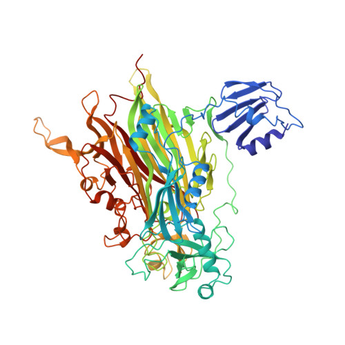
The crystal structure of the complex between the copper amine oxidase from Escherichia coli (ECAO) and a covalently bound inhibitor, 2-hydrazinopyridine, has been determined to a resolution of 2.0 A. The inhibitor covalently binds at the 5 position of the quinone ring of the cofactor, 2,4,5-trihydroxyphenylalaninequinone (TPQ). The inhibitor complex is analogous to the substrate Schiff base formed during the reaction with natural monoamine substrate. A proton is abstracted from a methylene group adjacent to the amine group by a catalytic base during the reaction. The inhibitor, however, has a nitrogen at this position, preventing proton abstraction and trapping the enzyme in a covalent complex. The electron density shows this nitrogen is hydrogen bonded to the side chain of Asp383, a totally conserved residue, identifying it as the probable catalytic base. The positioning of Asp383 is such that the pro-S proton of a substrate would be abstracted, consistent with the stereospecificity of the enzyme determined by 1H NMR spectroscopy. Site-directed mutagenesis and in vivo suppression have been used to substitute Asp383 for 12 other residues. The resulting proteins either lack or, in the case of glutamic acid, have very low enzyme activity consistent with an essential catalytic role for Asp383. The O4 position on the quinone ring is involved in a short hydrogen bond with the hydroxyl of conserved residue Tyr369. The distance between the oxygens is less than 2.5 A, consistent with a shared proton, and suggesting ionization at the O4 position of the quinone ring. The Tyr369 residue appears to play an important role in stabilizing the position of the quinone/inhibitor complex. The O2 position on the quinone ring is hydrogen bonded to the apical water ligand of the copper. The basal water ligand, which lies 2.0 A from the copper in the native structure, is at a distance of 3.0 A in the complex. In the native structure, the active site is completely buried, with no obvious route for entry of substrate. In the complex, the tip of the pyridine ring of the bound inhibitor is on the surface of the protein at the edge of the interface between domains 3 and 4, suggesting this as the entry point for the amine substrate.
- Department of Biochemistry and Molecular Biology, University of Leeds, United Kingdom.
Organizational Affiliation: