1S8L

Anion-free form of the D85S mutant of bacteriorhodopsin from crystals grown in the presence of halide


Experimental Data Snapshot

  • Method: X-RAY DIFFRACTION
  • Resolution: 2.30 Å
  • R-Value Free: 0.253 
  • R-Value Work: 0.221 
  • R-Value Observed: 0.221 

wwPDB Validation   3D Report Full Report


Ligand Structure Quality Assessment 


This is version 1.4 of the entry. See complete history


Literature

Specificity of anion binding in the substrate pocket of bacteriorhodopsin.

Facciotti, M.T.Cheung, V.S.Lunde, C.S.Rouhani, S.Baliga, N.S.Glaeser, R.M.

(2004) Biochemistry 43: 4934-4943

  • DOI: https://doi.org/10.1021/bi035757s
  • Primary Citation of Related Structures:  
    1S8J, 1S8L

  • PubMed Abstract: 

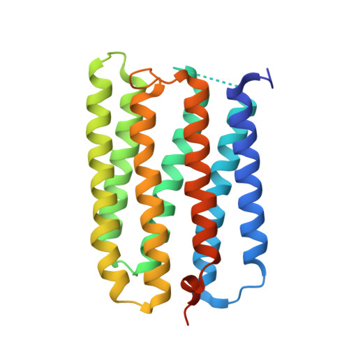
    The structure of the D85S mutant of bacteriorhodopsin with a nitrate anion bound in the Schiff base binding site and the structure of the anion-free protein have been obtained in the same crystal form. Together with the previously solved structures of this anion pump, in both the anion-free state and bromide-bound state, these new structures provide insight into how this mutant of bacteriorhodopsin is able to bind a variety of different anions in the same binding pocket. The structural analysis reveals that the main structural change that accommodates different anions is the repositioning of the polar side chain of S85. On the basis of these X-ray crystal structures, the prediction is then made that the D85S/D212N double mutant might bind similar anions and do so over a broader pH range than does the single mutant. Experimental comparison of the dissociation constants, K(d), for a variety of anions confirms this prediction and demonstrates, in addition, that the binding affinity is dramatically improved by the D212N substitution.


  • Organizational Affiliation

    Life Sciences Division, Lawrence Berkeley National Laboratory, Berkeley, California 94720, USA.


Macromolecules
Find similar proteins by:  (by identity cutoff)  |  3D Structure
Entity ID: 1
MoleculeChains Sequence LengthOrganismDetailsImage
Bacteriorhodopsin precursor249Halobacterium sp.Mutation(s): 1 
Gene Names: BOPVNG1467G
Membrane Entity: Yes 
UniProt
Find proteins for P02945 (Halobacterium salinarum (strain ATCC 700922 / JCM 11081 / NRC-1))
Explore P02945 
Go to UniProtKB:  P02945
Entity Groups  
Sequence Clusters30% Identity50% Identity70% Identity90% Identity95% Identity100% Identity
UniProt GroupP02945
Sequence Annotations
Expand
  • Reference Sequence
Small Molecules
Ligands 2 Unique
IDChains Name / Formula / InChI Key2D Diagram3D Interactions
LI1
Query on LI1

Download Ideal Coordinates CCD File 
C [auth A]
D [auth A]
E [auth A]
F [auth A]
G [auth A]
C [auth A],
D [auth A],
E [auth A],
F [auth A],
G [auth A],
H [auth A],
I [auth A],
J [auth A]
1-[2,6,10.14-TETRAMETHYL-HEXADECAN-16-YL]-2-[2,10,14-TRIMETHYLHEXADECAN-16-YL]GLYCEROL
C42 H86 O3
YERVUJAKCNBGCR-BIHSMRAKSA-N
RET
Query on RET

Download Ideal Coordinates CCD File 
B [auth A]RETINAL
C20 H28 O
NCYCYZXNIZJOKI-OVSJKPMPSA-N
Experimental Data & Validation

Experimental Data

  • Method: X-RAY DIFFRACTION
  • Resolution: 2.30 Å
  • R-Value Free: 0.253 
  • R-Value Work: 0.221 
  • R-Value Observed: 0.221 
  • Space Group: C 2 2 21
Unit Cell:
Length ( Å )Angle ( ˚ )
a = 52.725α = 90
b = 121.335β = 90
c = 74.204γ = 90
Software Package:
Software NamePurpose
CNSrefinement
MOSFLMdata reduction
CCP4data scaling
CNSphasing

Structure Validation

View Full Validation Report


Ligand Structure Quality Assessment 


Entry History 

Deposition Data

Revision History  (Full details and data files)

  • Version 1.0: 2004-06-08
    Type: Initial release
  • Version 1.1: 2008-04-29
    Changes: Version format compliance
  • Version 1.2: 2011-07-13
    Changes: Version format compliance
  • Version 1.3: 2021-10-27
    Changes: Database references, Derived calculations
  • Version 1.4: 2023-08-23
    Changes: Data collection, Refinement description