1S1Q

TSG101(UEV) domain in complex with Ubiquitin


Experimental Data Snapshot

  • Method: X-RAY DIFFRACTION
  • Resolution: 2.00 Å
  • R-Value Free: 0.240 
  • R-Value Work: 0.201 
  • R-Value Observed: 0.203 

wwPDB Validation   3D Report Full Report


This is version 1.2 of the entry. See complete history


Literature

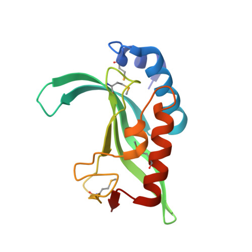
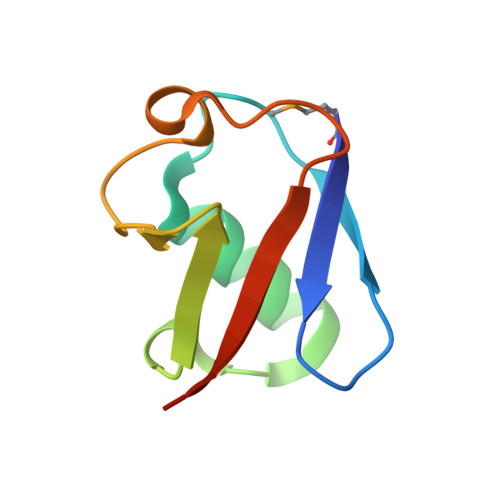
Ubiquitin recognition by the human TSG101 protein

Sundquist, W.I.Schubert, H.L.Kelly, B.N.Hill, G.C.Holton, J.M.Hill, C.P.

(2004) Mol Cell 13: 783-789

  • DOI: https://doi.org/10.1016/s1097-2765(04)00129-7
  • Primary Citation of Related Structures:  
    1S1Q

  • PubMed Abstract: 
  • The UEV domain of the TSG101 protein functions in both HIV-1 budding and the vacuolar protein sorting (VPS) pathway, where it binds ubiquitylated proteins as they are sorted into vesicles that bud into late endosomal compartments called multivesicular bodies (MVBs) ...

    The UEV domain of the TSG101 protein functions in both HIV-1 budding and the vacuolar protein sorting (VPS) pathway, where it binds ubiquitylated proteins as they are sorted into vesicles that bud into late endosomal compartments called multivesicular bodies (MVBs). TSG101 UEV-ubiquitin interactions are therefore important for delivery of both substrates and hydrolytic enzymes to lysosomes, which receive proteins via fusion with MVBs. Here, we report the crystal structure of the TSG101 UEV domain in complex with ubiquitin at 2.0 A resolution. TSG101 UEV contacts the Ile44 surface and an adjacent loop of ubiquitin through a highly solvated interface. Mutations that disrupt the interface inhibit MVB sorting, and the structure also explains how the TSG101 UEV can independently bind its ubiquitin and Pro-Thr/Ser-Ala-Pro peptide ligands. Remarkably, comparison with mapping data from other UEV and related E2 proteins indicates that although the different E2/UEV domains share the same structure and have conserved ubiquitin binding activity, they bind through very different interfaces.


    Organizational Affiliation

    Department of Biochemistry, University of Utah, Salt Lake City, UT 84132, USA. wes@biochem.utah.edu


Macromolecules
Find similar proteins by:  (by identity cutoff)  |  3D Structure
Entity ID: 1
MoleculeChainsSequence LengthOrganismDetailsImage
Tumor susceptibility gene 101 proteinA, C145Homo sapiensMutation(s): 5 
Gene Names: TSG101
UniProt & NIH Common Fund Data Resources
Find proteins for Q99816 (Homo sapiens)
Explore Q99816 
Go to UniProtKB:  Q99816
PHAROS:  Q99816
Entity Groups  
Sequence Clusters30% Identity50% Identity70% Identity90% Identity95% Identity100% Identity
UniProt GroupQ99816
Protein Feature View
Expand
  • Reference Sequence
Find similar proteins by:  (by identity cutoff)  |  3D Structure
Entity ID: 2
MoleculeChainsSequence LengthOrganismDetailsImage
ubiquitinB, D76Homo sapiensMutation(s): 1 
Gene Names: UBC
UniProt & NIH Common Fund Data Resources
Find proteins for P0CG48 (Homo sapiens)
Explore P0CG48 
Go to UniProtKB:  P0CG48
PHAROS:  P0CG48
Entity Groups  
Sequence Clusters30% Identity50% Identity70% Identity90% Identity95% Identity100% Identity
UniProt GroupP0CG48
Protein Feature View
Expand
  • Reference Sequence
Experimental Data & Validation

Experimental Data

  • Method: X-RAY DIFFRACTION
  • Resolution: 2.00 Å
  • R-Value Free: 0.240 
  • R-Value Work: 0.201 
  • R-Value Observed: 0.203 
  • Space Group: C 1 2 1
Unit Cell:
Length ( Å )Angle ( ˚ )
a = 143.634α = 90
b = 59.154β = 128.67
c = 93.998γ = 90
Software Package:
Software NamePurpose
DENZOdata reduction
SCALEPACKdata scaling
MOLREPphasing
REFMACrefinement

Structure Validation

View Full Validation Report


Entry History 

Deposition Data

Revision History  (Full details and data files)

  • Version 1.0: 2004-05-04
    Type: Initial release
  • Version 1.1: 2008-04-29
    Changes: Version format compliance
  • Version 1.2: 2011-07-13
    Changes: Derived calculations, Version format compliance