1QAE | pdb_00001qae

THE ACTIVE SITE OF SERRATIA ENDONUCLEASE CONTAINS A CONSERVED MAGNESIUM-WATER CLUSTER


Experimental Data Snapshot

  • Method: X-RAY DIFFRACTION
  • Resolution: 2.05 Å
  • R-Value Free: 
    0.210 (Depositor) 
  • R-Value Work: 
    0.167 (Depositor) 

Starting Model: experimental
View more details

wwPDB Validation   3D Report Full Report


This is version 1.6 of the entry. See complete history


Literature

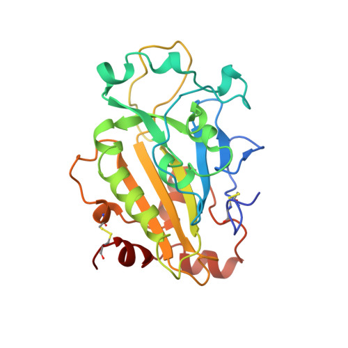
The active site of Serratia endonuclease contains a conserved magnesium-water cluster.

Miller, M.D.Cai, J.Krause, K.L.

(1999) J Mol Biology 288: 975-988

  • DOI: https://doi.org/10.1006/jmbi.1999.2729
  • Primary Citation of Related Structures:  
    1QAE

  • PubMed Abstract: 

    Serratia endonuclease is an important member of a class of magnesium dependent nucleases that are widely distributed in nature. Here, we describe the location and geometry of a magnesium-water cluster within the active site of this enzyme. The sole protein ligand of the magnesium atom is Asn119; this metal ion is also associated with five water molecules to complete an octahedral coordination complex. These water molecules are very well ordered and there is no evidence of rotational disorder or motion. Glu127 and His89 are located nearby and each is hydrogen bonded to water molecules in the coordination sphere. Asp86 is not chelated to the magnesium or its surrounding water molecules. Results of kinetics and site-specific mutagenesis experiments suggest that this metal-water cluster contains the catalytic metal ion of this enzyme. All residues which hydrogen bond to the water molecules that coordinate the magnesium atom are conserved in nucleases homologous to Serratia endonuclease, suggesting that the water cluster is a conserved feature of this family of enzymes. We offer a detailed structural comparison to one other nuclease, the homing endonuclease I-PpoI, that has recently been shown, in spite of a lack of sequence homology, to share a similar active site geometry to Serratia endonuclease. Evidence from both of these structures suggests that the magnesium of Serratia nuclease participates in catalysis via an inner sphere mechanism.


  • Organizational Affiliation
    • Department of Biology and Biochemistry, University of Houston, Houston, TX, 77204-5934, USA.

Macromolecules
Find similar proteins by:  (by identity cutoff)  |  3D Structure
Entity ID: 1
MoleculeChains Sequence LengthOrganismDetailsImage
PROTEIN (EXTRACELLULAR ENDONUCLEASE)
A, B
245Serratia marcescensMutation(s): 0 
EC: 3.1.30.2
UniProt
Find proteins for P13717 (Serratia marcescens)
Explore P13717 
Go to UniProtKB:  P13717
Entity Groups  
Sequence Clusters30% Identity50% Identity70% Identity90% Identity95% Identity100% Identity
UniProt GroupP13717
Sequence Annotations
Expand
  • Reference Sequence
Experimental Data & Validation

Experimental Data

  • Method: X-RAY DIFFRACTION
  • Resolution: 2.05 Å
  • R-Value Free:  0.210 (Depositor) 
  • R-Value Work:  0.167 (Depositor) 
Space Group: P 21 21 2
Unit Cell:
Length ( Å )Angle ( ˚ )
a = 106.7α = 90
b = 74.5β = 90
c = 68.9γ = 90
Software Package:
Software NamePurpose
MADNESSdata collection
PROCORdata scaling
PROCORdata reduction
XSCALEdata scaling
ARP/wARPmodel building
X-PLORmodel building
X-PLORrefinement
MADNESSdata reduction
X-PLORphasing

Structure Validation

View Full Validation Report


Entry History 

Deposition Data

Revision History  (Full details and data files)

  • Version 1.0: 1999-05-18
    Type: Initial release
  • Version 1.1: 2007-10-16
    Changes: Version format compliance
  • Version 1.2: 2011-07-13
    Changes: Version format compliance
  • Version 1.3: 2017-10-04
    Changes: Refinement description
  • Version 1.4: 2018-04-18
    Changes: Data collection
  • Version 1.5: 2024-04-03
    Changes: Data collection, Database references, Derived calculations, Refinement description
  • Version 1.6: 2024-10-16
    Changes: Structure summary