1NZS

NMR structures of phosphorylated carboxy terminus of bovine rhodopsin in arrestin-bound state


Experimental Data Snapshot

  • Method: SOLUTION NMR
  • Conformers Calculated: 100 
  • Conformers Submitted: 16 
  • Selection Criteria: structures with the lowest energy 

wwPDB Validation   3D Report Full Report


This is version 1.3 of the entry. See complete history


Literature

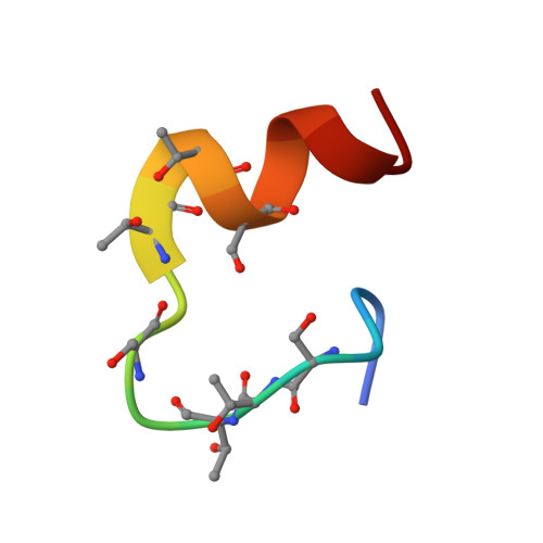
The arrestin-bound conformation and dynamics of the phosphorylated carboxy-terminal region of rhodopsin.

Kisselev, O.G.McDowell, J.H.Hargrave, P.A.

(2004) FEBS Lett 564: 307-311

  • DOI: https://doi.org/10.1016/S0014-5793(04)00226-1
  • Primary Citation of Related Structures:  
    1NZS

  • PubMed Abstract: 
  • Visual arrestin binds to the phosphorylated carboxy-terminal region of rhodopsin to block interactions with transducin and terminate signaling in the rod photoreceptor cells. A synthetic seven-phospho-peptide from the C-terminal region of rhodopsin, Rh(330-348), has been shown to bind arrestin and mimic inhibition of signal transduction ...

    Visual arrestin binds to the phosphorylated carboxy-terminal region of rhodopsin to block interactions with transducin and terminate signaling in the rod photoreceptor cells. A synthetic seven-phospho-peptide from the C-terminal region of rhodopsin, Rh(330-348), has been shown to bind arrestin and mimic inhibition of signal transduction. In this study, we examine conformational changes in this synthetic peptide upon binding to arrestin by high-resolution proton nuclear magnetic resonance (NMR). We show that the peptide is completely disordered in solution, but becomes structured upon binding to arrestin. A control, unphosphorylated peptide that fails to bind to arrestin remains highly disordered. Specific NMR distance constraints are used to model the arrestin-bound conformation. The models suggest that the phosphorylated carboxy-terminal region of rhodopsin, Rh(330-348), undergoes significant conformational changes and becomes structured upon binding to arrestin.


    Organizational Affiliation

    Department of Ophthalmology and Biochemistry, Saint Louis University School of Medicine, 1755 S. Grand Blvd., St. Louis, MO 63104, USA. kisselev@slu.edu


Macromolecules

Find similar proteins by:  Sequence   |   3D Structure  

Entity ID: 1
MoleculeChainsSequence LengthOrganismDetailsImage
19-mer peptide fragment of RHODOPSIN19N/AMutation(s): 7 
UniProt
Find proteins for P02699 (Bos taurus)
Explore P02699 
Go to UniProtKB:  P02699
Entity Groups  
Sequence Clusters30% Identity50% Identity70% Identity90% Identity95% Identity100% Identity
UniProt GroupP02699
Protein Feature View
Expand
  • Reference Sequence
Small Molecules
Modified Residues  2 Unique
IDChainsTypeFormula2D DiagramParent
SEP
Query on SEP
A
L-PEPTIDE LINKINGC3 H8 N O6 PSER
TPO
Query on TPO
A
L-PEPTIDE LINKINGC4 H10 N O6 PTHR
Experimental Data & Validation

Experimental Data

  • Method: SOLUTION NMR
  • Conformers Calculated: 100 
  • Conformers Submitted: 16 
  • Selection Criteria: structures with the lowest energy 

Structure Validation

View Full Validation Report


Entry History 

Deposition Data

Revision History  (Full details and data files)

  • Version 1.0: 2004-03-02
    Type: Initial release
  • Version 1.1: 2008-04-29
    Changes: Version format compliance
  • Version 1.2: 2011-07-13
    Changes: Version format compliance
  • Version 1.3: 2022-02-23
    Changes: Data collection, Database references, Derived calculations