1NAA | pdb_00001naa

Cellobiose Dehydrogenase Flavoprotein Fragment in Complex with Cellobionolactam


Experimental Data Snapshot

  • Method: X-RAY DIFFRACTION
  • Resolution: 1.80 Å
  • R-Value Free: 
    0.185 (Depositor), 0.204 (DCC) 
  • R-Value Work: 
    0.146 (Depositor), 0.174 (DCC) 
  • R-Value Observed: 
    0.146 (Depositor) 

Starting Model: experimental
View more details

wwPDB Validation 3D Report Full Report

Validation slider image for 1NAA

Ligand Structure Quality Assessment 


This is version 2.2 of the entry. See complete history

Literature

Mechanism of the reductive half-reaction in cellobiose dehydrogenase

Hallberg, B.M.Henriksson, G.Pettersson, G.Vasella, A.Divne, C.

(2003) J Biological Chem 278: 7160-7166

  • DOI: https://doi.org/10.1074/jbc.M210961200
  • Primary Citation Related Structures: 
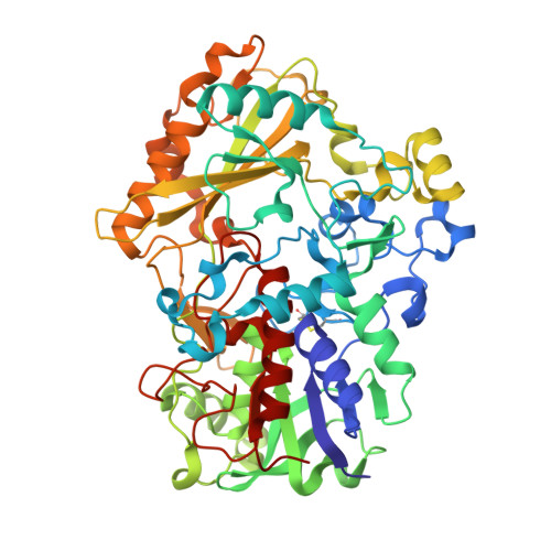
    1NAA

  • PubMed Abstract: 

    The extracellular flavocytochrome cellobiose dehydrogenase (CDH; EC ) participates in lignocellulose degradation by white-rot fungi with a proposed role in the early events of wood degradation. The complete hemoflavoenzyme consists of a catalytically active dehydrogenase fragment (DH(cdh)) connected to a b-type cytochrome domain via a linker peptide. In the reductive half-reaction, DH(cdh) catalyzes the oxidation of cellobiose to yield cellobiono-1,5-lactone. The active site of DH(cdh) is structurally similar to that of glucose oxidase and cholesterol oxidase, with a conserved histidine residue positioned at the re face of the flavin ring close to the N5 atom. The mechanisms of oxidation in glucose oxidase and cholesterol oxidase are still poorly understood, partly because of lack of experimental structure data or difficulties in interpreting existing data for enzyme-ligand complexes. Here we report the crystal structure of the Phanerochaete chrysosporium DH(cdh) with a bound inhibitor, cellobiono-1,5-lactam, at 1.8-A resolution. The distance between the lactam C1 and the flavin N5 is only 2.9 A, implying that in an approximately planar transition state, the maximum distance for the axial 1-hydrogen to travel for covalent addition to N5 is 0.8-0.9 A. The lactam O1 interacts intimately with the side chains of His-689 and Asn-732. Our data lend substantial structural support to a reaction mechanism where His-689 acts as a general base by abstracting the O1 hydroxyl proton in concert with transfer of the C1 hydrogen as hydride to the re face of the flavin N5.


  • Organizational Affiliation
    • Department of Biotechnology, Albanova University Center, KTH, SE-106 91 Stockholm, Sweden.

Macromolecule Content 

  • Total Structure Weight: 118.48 kDa 
  • Atom Count: 9,365 
  • Modeled Residue Count: 1,082 
  • Deposited Residue Count: 1,082 
  • Unique protein chains: 1

Macromolecules

Find similar proteins by:|  3D Structure
Entity ID: 1
MoleculeChains  Sequence LengthOrganismDetailsImage
Cellobiose dehydrogenase
A, B
541Phanerodontia chrysosporiumMutation(s): 0 
EC: 1.1.99.18
UniProt
Find proteins for Q01738 (Phanerodontia chrysosporium)
Explore Q01738 
Go to UniProtKB:  Q01738
Entity Groups
Sequence Clusters30% Identity50% Identity70% Identity90% Identity95% Identity100% Identity
UniProt GroupQ01738
Glycosylation
Glycosylation Sites: 2
Sequence Annotations
Expand
Reference Sequence

Small Molecules

Ligands 3 Unique
IDChains Name / Formula / InChI Key2D Diagram3D Interactions
6FA

Query on 6FA


Download:Ideal Coordinates CCD File
F [auth A],
J [auth B]
6-HYDROXY-FLAVIN-ADENINE DINUCLEOTIDE
C27 H33 N9 O16 P2
BJSUUWFQAMLNKU-OKXKTURISA-N
ABL

Query on ABL


Download:Ideal Coordinates CCD File
G [auth A],
K [auth B]
(2R,3R,4R,5R)-4,5-dihydroxy-2-(hydroxymethyl)-6-oxopiperidin-3-yl beta-D-glucopyranoside
C12 H21 N O10
WXSNJJDPPISYEF-ZNLUKOTNSA-N
NAG

Query on NAG


Download:Ideal Coordinates CCD File
C [auth A],
D [auth A],
E [auth A],
H [auth B],
I [auth B]
2-acetamido-2-deoxy-beta-D-glucopyranose
C8 H15 N O6
OVRNDRQMDRJTHS-FMDGEEDCSA-N

Experimental Data & Validation

Experimental Data

  • Method: X-RAY DIFFRACTION
  • Resolution: 1.80 Å
  • R-Value Free:  0.185 (Depositor), 0.204 (DCC) 
  • R-Value Work:  0.146 (Depositor), 0.174 (DCC) 
  • R-Value Observed: 0.146 (Depositor) 
Space Group: H 3
Unit Cell:
Length ( Å )Angle ( ˚ )
a = 185.952α = 90
b = 185.952β = 90
c = 81.438γ = 120
Software Package:
Software NamePurpose
REFMACrefinement
SCALAdata scaling
CNSrefinement
MOSFLMdata reduction
CCP4data scaling
CNSphasing

Structure Validation

View Full Validation Report


Ligand Structure Quality Assessment 


Entry History 

Deposition Data

Revision History  (Full details and data files)

  • Version 1.0: 2003-01-14
    Type: Initial release
  • Version 1.1: 2008-04-28
    Changes: Version format compliance
  • Version 1.2: 2011-07-13
    Changes: Non-polymer description, Version format compliance
  • Version 1.3: 2014-11-12
    Changes: Structure summary
  • Version 1.4: 2019-07-24
    Changes: Data collection, Derived calculations, Refinement description
  • Version 2.0: 2020-07-29
    Type: Remediation
    Reason: Carbohydrate remediation
    Changes: Atomic model, Data collection, Derived calculations, Structure summary
  • Version 2.1: 2023-08-16
    Changes: Data collection, Database references, Refinement description, Structure summary
  • Version 2.2: 2024-11-20
    Changes: Structure summary