1MAB

RAT LIVER F1-ATPASE


Experimental Data Snapshot

  • Method: X-RAY DIFFRACTION
  • Resolution: 2.80 Å
  • R-Value Free: 0.290 
  • R-Value Work: 0.217 
  • R-Value Observed: 0.217 

wwPDB Validation   3D Report Full Report


This is version 1.5 of the entry. See complete history


Literature

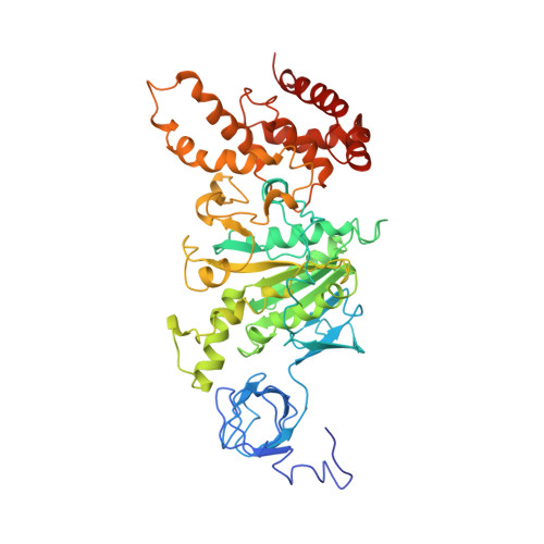
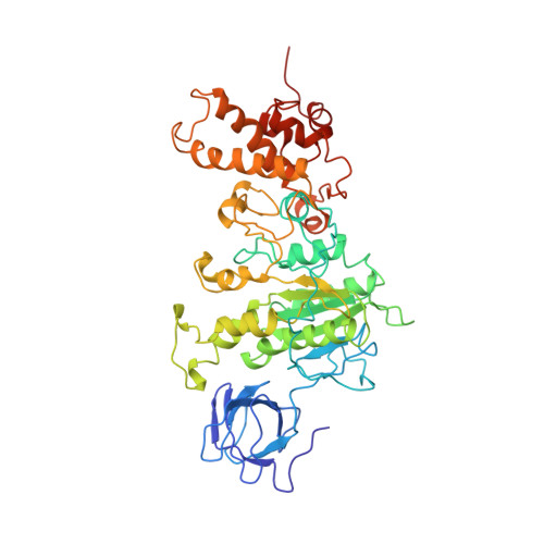
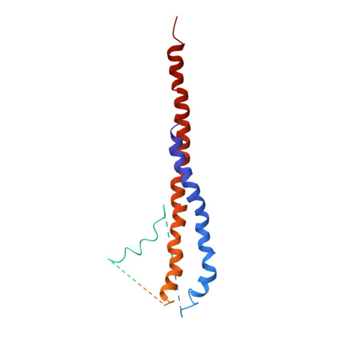
The 2.8-A structure of rat liver F1-ATPase: configuration of a critical intermediate in ATP synthesis/hydrolysis.

Bianchet, M.A.Hullihen, J.Pedersen, P.L.Amzel, L.M.

(1998) Proc Natl Acad Sci U S A 95: 11065-11070

  • DOI: https://doi.org/10.1073/pnas.95.19.11065
  • Primary Citation of Related Structures:  
    1MAB

  • PubMed Abstract: 

    During mitochondrial ATP synthesis, F1-ATPase-the portion of the ATP synthase that contains the catalytic and regulatory nucleotide binding sites-undergoes a series of concerted conformational changes that couple proton translocation to the synthesis of the high levels of ATP required for cellular function. In the structure of the rat liver F1-ATPase, determined to 2.8-A resolution in the presence of physiological concentrations of nucleotides, all three beta subunits contain bound nucleotide and adopt similar conformations. This structure provides the missing configuration of F1 necessary to define all intermediates in the reaction pathway. Incorporation of this structure suggests a mechanism of ATP synthesis/hydrolysis in which configurations of the enzyme with three bound nucleotides play an essential role.


  • Organizational Affiliation

    Department of Biophysics and Biophysical Chemistry, The Johns Hopkins University School of Medicine, 725 North Wolfe Street, Baltimore, MD 21205-2185, USA. mario@neruda.med.jhmi.edu


Macromolecules
Find similar proteins by:  (by identity cutoff)  |  3D Structure
Entity ID: 1
MoleculeChains Sequence LengthOrganismDetailsImage
PROTEIN (F1-ATPASE ALPHA CHAIN)510Rattus norvegicusMutation(s): 0 
UniProt
Find proteins for P15999 (Rattus norvegicus)
Explore P15999 
Go to UniProtKB:  P15999
Entity Groups  
Sequence Clusters30% Identity50% Identity70% Identity90% Identity95% Identity100% Identity
UniProt GroupP15999
Sequence Annotations
Expand
  • Reference Sequence
Find similar proteins by:  (by identity cutoff)  |  3D Structure
Entity ID: 2
MoleculeChains Sequence LengthOrganismDetailsImage
PROTEIN (F1-ATPASE BETA CHAIN)479Rattus norvegicusMutation(s): 0 
EC: 7.1.2.2
UniProt
Find proteins for P10719 (Rattus norvegicus)
Explore P10719 
Go to UniProtKB:  P10719
Entity Groups  
Sequence Clusters30% Identity50% Identity70% Identity90% Identity95% Identity100% Identity
UniProt GroupP10719
Sequence Annotations
Expand
  • Reference Sequence
Find similar proteins by:  (by identity cutoff)  |  3D Structure
Entity ID: 3
MoleculeChains Sequence LengthOrganismDetailsImage
PROTEIN (F1-ATPASE GAMMA CHAIN)C [auth G]270Rattus norvegicusMutation(s): 0 
UniProt
Find proteins for P35435 (Rattus norvegicus)
Explore P35435 
Go to UniProtKB:  P35435
Entity Groups  
Sequence Clusters30% Identity50% Identity70% Identity90% Identity95% Identity100% Identity
UniProt GroupP35435
Sequence Annotations
Expand
  • Reference Sequence
Small Molecules
Experimental Data & Validation

Experimental Data

  • Method: X-RAY DIFFRACTION
  • Resolution: 2.80 Å
  • R-Value Free: 0.290 
  • R-Value Work: 0.217 
  • R-Value Observed: 0.217 
  • Space Group: H 3 2
Unit Cell:
Length ( Å )Angle ( ˚ )
a = 143.67α = 90
b = 143.67β = 90
c = 361.15γ = 120
Software Package:
Software NamePurpose
X-PLORrefinement

Structure Validation

View Full Validation Report


Entry History 

Deposition Data

Revision History  (Full details and data files)

  • Version 1.0: 1998-09-30
    Type: Initial release
  • Version 1.1: 2008-04-27
    Changes: Version format compliance
  • Version 1.2: 2011-07-13
    Changes: Version format compliance
  • Version 1.3: 2017-10-04
    Changes: Refinement description
  • Version 1.4: 2023-05-31
    Changes: Database references, Derived calculations
  • Version 1.5: 2024-05-22
    Changes: Data collection