A new peculiar DNA structure: NMR solution structure of a DNA kissing complex.
Barbault, F., Huynh-Dinh, T., Paoletti, J., Lancelot, G.(2002) J Biomol Struct Dyn 19: 649-658
- PubMed: 11843626 Search on PubMed
- DOI: https://doi.org/10.1080/07391102.2002.10506771
- Primary Citation Related Structures:
1JU0, 1JUA - PubMed Abstract:
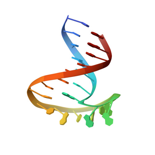
The deoxyoligoribonucleotide d(CTTGCTGAAGCGCGCACGGCAAG) (dSL1) corresponding to the reverse transcripted sequence of the dimerization initiation site SL1 of HIV- 1(Lai) RNA was synthesized using phosphoramidite chemistry. Like its oligoribonucleotide counterpart, dSL1 dimerized spontaneously in solution. Here we report the first NMR solution structure of a kissing complex formed with two DNA strands. The melting point of the DNA dimer (35 degrees C) was found slightly higher than the one of the corresponding RNA dimer (32 degrees C). Despite this only slight difference in melting point, several structural differences were observed between the ribo- and the deoxyribo- dimers. The solution structure of the deoxy- dimer was a symmetric homodimer with a loop-loop interaction stabilized by four central G-C base-pairs, a head to tail A-A base-pair arrangement between the A8 residues of the two strands and a stacking of A9 with C15. As a consequence, G10 was not paired and occupied a position outside the stem and the loop. Each stem was formed by seven base-pairs whose axis made an angle of about 100 degree with the plane of the loops. The distortion of the helix at the junction of the stem and of the loop induced a fold up of the A8pA9 step with a phosphate-phosphate distance lowered to 4.5 A. The plane of the non-canonical A-A base-pair was oriented perpendicularly to the axis of the stems. The four central base-pairs formed an open fan-shaped motif with an angle of 20 degrees between the bases and each of them was oriented perpendicularly to the A8-A8 plane. The deviation of the computed chemical shifts and the experimental ones for the aromatic proton was always less than 0.25ppm for each of the 16 converged solution structures and their average less than 0.1ppm.
- Centre de Biophysique Moléculaire, Rue Charles Sadron, 45071 Orléans Cedex 02, France.
Organizational Affiliation: