1INU

CRYSTAL STRUCTURE OF MUTANT HUMAN LYSOZYME SUBSTITUTED AT THE SURFACE POSITIONS


Experimental Data Snapshot

  • Method: X-RAY DIFFRACTION
  • Resolution: 1.80 Å
  • R-Value Work: 0.169 
  • R-Value Observed: 0.169 

wwPDB Validation   3D Report Full Report


This is version 1.5 of the entry. See complete history


Literature

Positive contribution of hydration structure on the surface of human lysozyme to the conformational stability.

Funahashi, J.Takano, K.Yamagata, Y.Yutani, K.

(2002) J Biol Chem 277: 21792-21800

  • DOI: https://doi.org/10.1074/jbc.M110728200
  • Primary Citation of Related Structures:  
    1GF8, 1GF9, 1GFA, 1GFE, 1GFG, 1GFH, 1GFJ, 1GFK, 1GFR, 1GFT, 1GFU, 1GFV, 1INU

  • PubMed Abstract: 

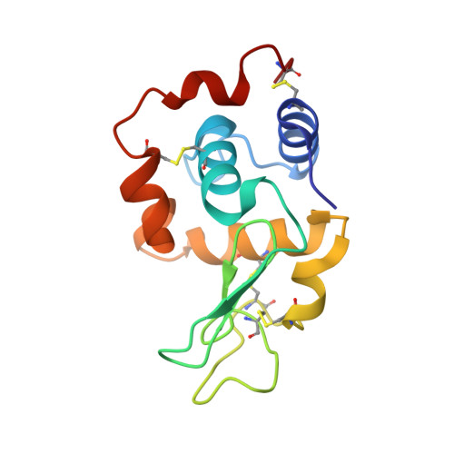
    Water molecules make a hydration structure with the network of hydrogen bonds, covering on the surface of proteins. To quantitatively estimate the contribution of the hydration structure to protein stability, a series of hydrophilic mutant human lysozymes (Val to Ser, Tyr, Asp, Asn, and Arg) modified at three different positions on the surface, which are located in the alpha-helix (Val-110), the beta-sheet (Val-2), and the loop (Val-74), were constructed. Their thermodynamic parameters of denaturation and crystal structures were examined by calorimetry and by x-ray crystallography at 100 K, respectively. The introduced polar residues made hydrogen bonds with protein atoms and/or water molecules, sometimes changing the hydration structure around the mutation site. Changes in the stability of the mutant proteins can be evaluated by a unique equation that considers the conformational changes resulting from the substitutions. Using this analysis, the relationship between the changes in the stabilities and the hydration structures for mutant human lysozymes substituted on the surface could be quantitatively estimated. The analysis indicated that the hydration structure on protein surface plays an important role in determining the conformational stability of the protein.


  • Organizational Affiliation

    Institute for Protein Research, Osaka University, Yamadaoka, Suita, Osaka 565-0871, Japan.


Macromolecules
Find similar proteins by:  (by identity cutoff)  |  3D Structure
Entity ID: 1
MoleculeChains Sequence LengthOrganismDetailsImage
LYSOZYME130Homo sapiensMutation(s): 1 
EC: 3.2.1.17
UniProt & NIH Common Fund Data Resources
Find proteins for P61626 (Homo sapiens)
Explore P61626 
Go to UniProtKB:  P61626
PHAROS:  P61626
GTEx:  ENSG00000090382 
Entity Groups  
Sequence Clusters30% Identity50% Identity70% Identity90% Identity95% Identity100% Identity
UniProt GroupP61626
Sequence Annotations
Expand
  • Reference Sequence
Small Molecules
Ligands 1 Unique
IDChains Name / Formula / InChI Key2D Diagram3D Interactions
NA
Query on NA

Download Ideal Coordinates CCD File 
B [auth A]SODIUM ION
Na
FKNQFGJONOIPTF-UHFFFAOYSA-N
Experimental Data & Validation

Experimental Data

  • Method: X-RAY DIFFRACTION
  • Resolution: 1.80 Å
  • R-Value Work: 0.169 
  • R-Value Observed: 0.169 
  • Space Group: P 21 21 21
Unit Cell:
Length ( Å )Angle ( ˚ )
a = 56.45α = 90
b = 60.68β = 90
c = 33.06γ = 90
Software Package:
Software NamePurpose
X-PLORmodel building
X-PLORrefinement
X-PLORphasing

Structure Validation

View Full Validation Report


Entry History 

Deposition Data

Revision History  (Full details and data files)

  • Version 1.0: 2000-12-20
    Type: Initial release
  • Version 1.1: 2008-04-27
    Changes: Version format compliance
  • Version 1.2: 2011-07-13
    Changes: Version format compliance
  • Version 1.3: 2017-10-04
    Changes: Refinement description
  • Version 1.4: 2021-11-10
    Changes: Database references, Derived calculations
  • Version 1.5: 2023-12-27
    Changes: Data collection