1I44 | pdb_00001i44

CRYSTALLOGRAPHIC STUDIES OF AN ACTIVATION LOOP MUTANT OF THE INSULIN RECEPTOR TYROSINE KINASE


Experimental Data Snapshot

  • Method: X-RAY DIFFRACTION
  • Resolution: 2.40 Å
  • R-Value Free: 
    0.268 (Depositor) 
  • R-Value Work: 
    0.210 (Depositor), 0.221 (DCC) 
  • R-Value Observed: 
    0.210 (Depositor) 

Starting Model: experimental
View more details

wwPDB Validation 3D Report Full Report

Validation slider image for 1I44

Ligand Structure Quality Assessment 


This is version 1.4 of the entry. See complete history

Literature

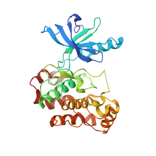
Crystallographic and solution studies of an activation loop mutant of the insulin receptor tyrosine kinase: insights into kinase mechanism.

Till, J.H.Ablooglu, A.J.Frankel, M.Bishop, S.M.Kohanski, R.A.Hubbard, S.R.

(2001) J Biological Chem 276: 10049-10055

  • DOI: https://doi.org/10.1074/jbc.M010161200
  • Primary Citation Related Structures: 
    1I44

  • PubMed Abstract: 

    The tyrosine kinase domain of the insulin receptor is subject to autoinhibition in the unphosphorylated basal state via steric interactions involving the activation loop. A mutation in the activation loop designed to relieve autoinhibition, Asp-1161 --> Ala, substantially increases the ability of the unphosphorylated kinase to bind ATP. The crystal structure of this mutant in complex with an ATP analog has been determined at 2.4-A resolution. The structure shows that the active site is unobstructed, but the end of the activation loop is disordered and therefore the binding site for peptide substrates is not fully formed. In addition, Phe-1151 of the protein kinase-conserved DFG motif, at the beginning of the activation loop, hinders closure of the catalytic cleft and proper positioning of alpha-helix C for catalysis. These results, together with viscometric kinetic measurements, suggest that peptide substrate binding induces a reconfiguration of the unphosphorylated activation loop prior to the catalytic step. The crystallographic and solution studies provide new insights into the mechanism by which the activation loop controls phosphoryl transfer as catalyzed by the insulin receptor.


  • Organizational Affiliation
    • Skirball Institute of Biomolecular Medicine and Department of Pharmacology, New York University School of Medicine, New York, New York 10016, USA.

Macromolecule Content 

  • Total Structure Weight: 35.28 kDa 
  • Atom Count: 2,378 
  • Modeled Residue Count: 282 
  • Deposited Residue Count: 306 
  • Unique protein chains: 1

Macromolecules

Find similar proteins by:|  3D Structure
Entity ID: 1
MoleculeChains  Sequence LengthOrganismDetailsImage
INSULIN RECEPTOR306Homo sapiensMutation(s): 3 
EC: 2.7.1.112 (PDB Primary Data), 2.7.10.1 (UniProt)
UniProt & NIH Common Fund Data Resources
Find proteins for P06213 (Homo sapiens)
Explore P06213 
Go to UniProtKB:  P06213
PHAROS:  P06213
GTEx:  ENSG00000171105 
Entity Groups
Sequence Clusters30% Identity50% Identity70% Identity90% Identity95% Identity100% Identity
UniProt GroupP06213
Sequence Annotations
Expand
Reference Sequence

Small Molecules

Ligands 2 Unique
IDChains Name / Formula / InChI Key2D Diagram3D Interactions
ACP

Query on ACP


Download:Ideal Coordinates CCD File
C [auth A]PHOSPHOMETHYLPHOSPHONIC ACID ADENYLATE ESTER
C11 H18 N5 O12 P3
UFZTZBNSLXELAL-IOSLPCCCSA-N
MG

Query on MG


Download:Ideal Coordinates CCD File
B [auth A]MAGNESIUM ION
Mg
JLVVSXFLKOJNIY-UHFFFAOYSA-N

Experimental Data & Validation

Experimental Data

  • Method: X-RAY DIFFRACTION
  • Resolution: 2.40 Å
  • R-Value Free:  0.268 (Depositor) 
  • R-Value Work:  0.210 (Depositor), 0.221 (DCC) 
  • R-Value Observed: 0.210 (Depositor) 
Space Group: P 21 21 21
Unit Cell:
Length ( Å )Angle ( ˚ )
a = 57.857α = 90
b = 69.578β = 90
c = 89.26γ = 90
Software Package:
Software NamePurpose
AMoREphasing
CNSrefinement
DENZOdata reduction
SCALEPACKdata scaling

Structure Validation

View Full Validation Report


Ligand Structure Quality Assessment 


Entry History 

Deposition Data

Revision History  (Full details and data files)

  • Version 1.0: 2001-03-07
    Type: Initial release
  • Version 1.1: 2008-04-27
    Changes: Version format compliance
  • Version 1.2: 2011-07-13
    Changes: Version format compliance
  • Version 1.3: 2021-10-27
    Changes: Database references, Derived calculations
  • Version 1.4: 2023-08-09
    Changes: Data collection, Refinement description