1HBN | pdb_00001hbn

METHYL-COENZYME M REDUCTASE


Experimental Data Snapshot

  • Method: X-RAY DIFFRACTION
  • Resolution: 1.16 Å
  • R-Value Free: 
    0.190 (Depositor) 
  • R-Value Work: 
    0.124 (Depositor) 

wwPDB Validation 3D Report Full Report

Currently 1HBN does not have a validation slider image.


This is version 2.2 of the entry. See complete history

Literature

On the Mechanism of Biological Methane Formation: Structural Evidence for Conformational Changes in Methyl-Coenzyme M Reductase Upon Substrate Binding

Grabarse, W.Mahlert, F.Duin, E.C.Goubeaud, M.Shima, S.Thauer, R.K.Lamzin, V.Ermler, U.

(2001) J Mol Biology 309: 315

  • DOI: https://doi.org/10.1006/jmbi.2001.4647
  • Primary Citation Related Structures: 
    1HBM, 1HBN, 1HBO, 1HBU

  • PubMed Abstract: 

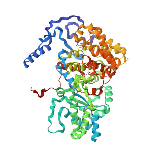
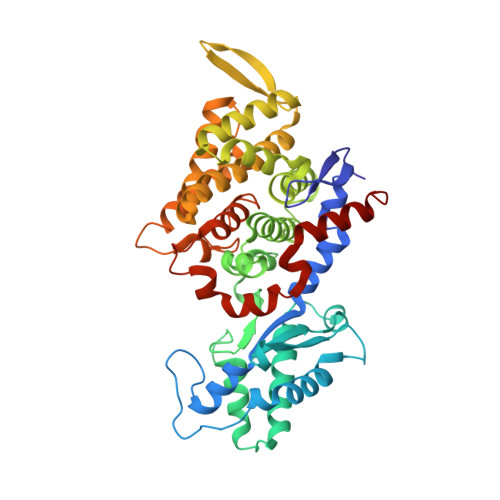
    Methyl-coenzyme M reductase (MCR) catalyzes the final reaction of the energy conserving pathway of methanogenic archaea in which methylcoenzyme M and coenzyme B are converted to methane and the heterodisulfide CoM-S-S-CoB. It operates under strictly anaerobic conditions and contains the nickel porphinoid F430 which is present in the nickel (I) oxidation state in the active enzyme. The known crystal structures of the inactive nickel (II) enzyme in complex with coenzyme M and coenzyme B (MCR-ox1-silent) and in complex with the heterodisulfide CoM-S-S-CoB (MCR-silent) were now refined at 1.16 A and 1.8 A resolution, respectively. The atomic resolution structure of MCR-ox1-silent describes the exact geometry of the cofactor F430, of the active site residues and of the modified amino acid residues. Moreover, the observation of 18 Mg2+ and 9 Na+ ions at the protein surface of the 300 kDa enzyme specifies typical constituents of binding sites for either ion. The MCR-silent and MCR-ox1-silent structures differed in the occupancy of bound water molecules near the active site indicating that a water chain is involved in the replenishment of the active site with water molecules. The structure of the novel enzyme state MCR-red1-silent at 1.8 A resolution revealed an active site only partially occupied by coenzyme M and coenzyme B. Increased flexibility and distinct alternate conformations were observed near the active site and the substrate channel. The electron density of the MCR-red1-silent state aerobically co-crystallized with coenzyme M displayed a fully occupied coenzyme M-binding site with no alternate conformations. Therefore, the structure was very similar to the MCR-ox1-silent state. As a consequence, the binding of coenzyme M induced specific conformational changes that postulate a molecular mechanism by which the enzyme ensures that methylcoenzyme M enters the substrate channel prior to coenzyme B as required by the active-site geometry. The three different enzymatically inactive enzyme states are discussed with respect to their enzymatically active precursors and with respect to the catalytic mechanism.


  • Organizational Affiliation
    • Max-Planck-Institut für Biophysik, Frankfurt, Germany.

Macromolecule Content 

  • Total Structure Weight: 277.25 kDa 
  • Atom Count: 21,958 
  • Modeled Residue Count: 2,474 
  • Deposited Residue Count: 2,478 
  • Unique protein chains: 3

Macromolecules

Find similar proteins by:|  3D Structure
Entity ID: 1
MoleculeChains  Sequence LengthOrganismDetailsImage
METHYL-COENZYME M REDUCTASE I ALPHA SUBUNIT
A, D
549Methanothermobacter thermautotrophicusMutation(s): 5 
EC: 2.8.4.1
UniProt
Find proteins for P11558 (Methanothermobacter marburgensis (strain ATCC BAA-927 / DSM 2133 / JCM 14651 / NBRC 100331 / OCM 82 / Marburg))
Explore P11558 
Go to UniProtKB:  P11558
Entity Groups
Sequence Clusters30% Identity50% Identity70% Identity90% Identity95% Identity100% Identity
UniProt GroupP11558
Sequence Annotations
Expand
Reference Sequence
Find similar proteins by:|  3D Structure
Entity ID: 2
MoleculeChains  Sequence LengthOrganismDetailsImage
METHYL-COENZYME M REDUCTASE I BETA SUBUNIT
B, E
442Methanothermobacter thermautotrophicusMutation(s): 0 
EC: 2.8.4.1
UniProt
Find proteins for P11560 (Methanothermobacter marburgensis (strain ATCC BAA-927 / DSM 2133 / JCM 14651 / NBRC 100331 / OCM 82 / Marburg))
Explore P11560 
Go to UniProtKB:  P11560
Entity Groups
Sequence Clusters30% Identity50% Identity70% Identity90% Identity95% Identity100% Identity
UniProt GroupP11560
Sequence Annotations
Expand
Reference Sequence
Find similar proteins by:|  3D Structure
Entity ID: 3
MoleculeChains  Sequence LengthOrganismDetailsImage
METHYL-COENZYME M REDUCTASE I GAMMA SUBUNIT
C, F
248Methanothermobacter thermautotrophicusMutation(s): 0 
EC: 2.8.4.1
UniProt
Find proteins for P11562 (Methanothermobacter marburgensis (strain ATCC BAA-927 / DSM 2133 / JCM 14651 / NBRC 100331 / OCM 82 / Marburg))
Explore P11562 
Go to UniProtKB:  P11562
Entity Groups
Sequence Clusters30% Identity50% Identity70% Identity90% Identity95% Identity100% Identity
UniProt GroupP11562
Sequence Annotations
Expand
Reference Sequence

Small Molecules

Ligands 8 Unique
IDChains Name / Formula / InChI Key2D Diagram3D Interactions
F43

Query on F43


Download:Ideal Coordinates CCD File
G [auth A],
HA [auth D]
FACTOR 430
C42 H51 N6 Ni O13
XLFIRMYGVLUNOY-SXMZNAGASA-M
TP7

Query on TP7


Download:Ideal Coordinates CCD File
H [auth A],
IA [auth D]
Coenzyme B
C11 H22 N O7 P S
JBJSVEVEEGOEBZ-SCZZXKLOSA-N
COM

Query on COM


Download:Ideal Coordinates CCD File
I [auth A],
JA [auth D]
1-THIOETHANESULFONIC ACID
C2 H6 O3 S2
ZNEWHQLOPFWXOF-UHFFFAOYSA-N
GOL

Query on GOL


Download:Ideal Coordinates CCD File
EA [auth C]
J [auth A]
K [auth A]
KA [auth D]
L [auth A]
EA [auth C],
J [auth A],
K [auth A],
KA [auth D],
L [auth A],
LA [auth D],
M [auth A],
MA [auth D],
NA [auth D],
TA [auth E],
Y [auth B]
GLYCEROL
C3 H8 O3
PEDCQBHIVMGVHV-UHFFFAOYSA-N
ZN

Query on ZN


Download:Ideal Coordinates CCD File
N [auth A]ZINC ION
Zn
PTFCDOFLOPIGGS-UHFFFAOYSA-N
CL

Query on CL


Download:Ideal Coordinates CCD File
AA [auth B],
UA [auth E]
CHLORIDE ION
Cl
VEXZGXHMUGYJMC-UHFFFAOYSA-M
MG

Query on MG


Download:Ideal Coordinates CCD File
AB [auth F]
BA [auth B]
BB [auth F]
DA [auth B]
FA [auth C]
AB [auth F],
BA [auth B],
BB [auth F],
DA [auth B],
FA [auth C],
GA [auth C],
O [auth A],
OA [auth D],
PA [auth D],
T [auth A],
U [auth A],
V [auth A],
VA [auth E],
W [auth A],
WA [auth E],
XA [auth E],
YA [auth E],
ZA [auth E]
MAGNESIUM ION
Mg
JLVVSXFLKOJNIY-UHFFFAOYSA-N
NA

Query on NA


Download:Ideal Coordinates CCD File
CA [auth B]
P [auth A]
Q [auth A]
QA [auth D]
R [auth A]
CA [auth B],
P [auth A],
Q [auth A],
QA [auth D],
R [auth A],
RA [auth D],
S [auth A],
SA [auth D],
X [auth A],
Z [auth B]
SODIUM ION
Na
FKNQFGJONOIPTF-UHFFFAOYSA-N
Modified Residues  5 Unique
IDChains TypeFormula2D DiagramParent
AGM
Query on AGM
A, D
L-PEPTIDE LINKINGC7 H17 N4 O2ARG
GL3
Query on GL3
A, D
L-PEPTIDE LINKINGC2 H5 N O SGLY
MGN
Query on MGN
A, D
L-PEPTIDE LINKINGC6 H12 N2 O3GLN
MHS
Query on MHS
A, D
L-PEPTIDE LINKINGC7 H11 N3 O2HIS
SMC
Query on SMC
A, D
L-PEPTIDE LINKINGC4 H9 N O2 SCYS

Experimental Data & Validation

Experimental Data

  • Method: X-RAY DIFFRACTION
  • Resolution: 1.16 Å
  • R-Value Free:  0.190 (Depositor) 
  • R-Value Work:  0.124 (Depositor) 
Space Group: P 1 21 1
Unit Cell:
Length ( Å )Angle ( ˚ )
a = 81.72α = 90
b = 116.883β = 92.02
c = 122.582γ = 90
Software Package:
Software NamePurpose
REFMACrefinement
DENZOdata reduction
SCALEPACKdata scaling
CCP4phasing

Structure Validation

View Full Validation Report

Currently 1HBN does not have a validation slider image.


Entry History 

Deposition Data

Revision History  (Full details and data files)

  • Version 1.0: 2001-08-16
    Type: Initial release
  • Version 1.1: 2015-05-20
    Changes: Derived calculations, Non-polymer description, Other, Version format compliance
  • Version 2.0: 2017-06-21
    Changes: Advisory, Atomic model
  • Version 2.1: 2019-07-24
    Changes: Data collection, Derived calculations
  • Version 2.2: 2025-04-09
    Changes: Advisory, Data collection, Database references, Derived calculations, Structure summary