Short Constrained Peptides that Inhibit HIV-1 Entry
Sia, S.K., Carr, P.A., Cochran, A.G., Malashkevich, V.M., Kim, P.S.(2002) Proc Natl Acad Sci U S A 99: 14664
- PubMed: 12417739 Search on PubMedSearch on PubMed Central
- DOI: https://doi.org/10.1073/pnas.232566599
- Primary Citation Related Structures:
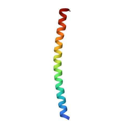
1GZL - PubMed Abstract:
Peptides corresponding to the C-terminal heptad repeat of HIV-1 gp41 (C-peptides) are potent inhibitors of HIV-1 entry into cells. Their mechanism of inhibition involves binding in a helical conformation to the central coiled coil of HIV-1 gp41 in a dominant-negative manner. Short C-peptides, however, have low binding affinity for gp41 and poor inhibitory activity, which creates an obstacle to the development of small drug-like C-peptides. To improve the inhibitory potency of short C-peptides that target the hydrophobic pocket region of gp41, we use two strategies to stabilize the C-peptide helix: chemical crosslinking and substitution with unnatural helix-favoring amino acids. In this study, the short linear peptide shows no significant inhibitory activity, but a constrained peptide (C14linkmid) inhibits cell-cell fusion at micromolar potency. Structural studies confirm that the constrained peptides bind to the gp41 hydrophobic pocket. Calorimetry reveals that, of the peptides analyzed, the most potent are those that best balance the changes in binding enthalpy and entropy, and surprisingly not those with the highest helical propensity as measured by circular dichroism spectroscopy. Our study reveals the thermodynamic basis of inhibition of an HIV C-peptide, demonstrates the utility of constraining methods for a short antiviral peptide inhibitor, and has implications for the future design of constrained peptides.
- Howard Hughes Medical Institute, Whitehead Institute for Biomedical Research, Department of Biology, Massachusetts Institute of Technology, Cambridge 02142, USA. sia@fas.harvard.edu
Organizational Affiliation: