Ras/Rap effector specificity determined by charge reversal.
Nassar, N., Horn, G., Herrmann, C., Block, C., Janknecht, R., Wittinghofer, A.(1996) Nat Struct Biol 3: 723-729
- PubMed: 8756332 Search on PubMed
- DOI: https://doi.org/10.1038/nsb0896-723
- Primary Citation Related Structures:
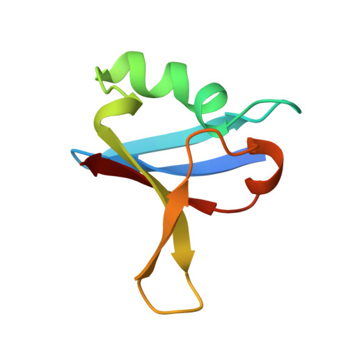
1GUA - PubMed Abstract:
Members of the Ras subfamily of small GTP-binding proteins have been shown to be promiscuous towards a variety of putative effector molecules such as the protein kinase c-Raf and the Ral-specific guanine nucleotide exchange factor (Ral-GEF). To address the question of specificity of interactions we have introduced the mutations E30D and K31E into Rap and show biochemically, by X-ray structure analysis and by transfection in vivo that the identical core effector region of Ras and Rap (residues 32-40) is responsible for molecular recognition, but that residues outside this region are responsible for the specificity of the interaction. The major determinant for the switch in specificity is the opposite charge of residue 31--Lys in Rap, Glu in Ras--which creates a favourable complementary interface for the Ras-Raf interaction.
- Max-Planck-Institut für molekulare Physiologie, Abteilung Strukturelle Biologie, Dortmund, Germany.
Organizational Affiliation: