Role of surface hydrophobic residues in the conformational stability of human lysozyme at three different positions.
Funahashi, J., Takano, K., Yamagata, Y., Yutani, K.(2000) Biochemistry 39: 14448-14456
- PubMed: 11087397 Search on PubMed
- DOI: https://doi.org/10.1021/bi0015717
- Primary Citation Related Structures:
1GAY, 1GB0, 1GB2, 1GB3, 1GB5, 1GB6, 1GB7, 1GB8, 1GB9, 1GBO, 1GBW, 1GBX, 1GBY, 1GBZ - PubMed Abstract:
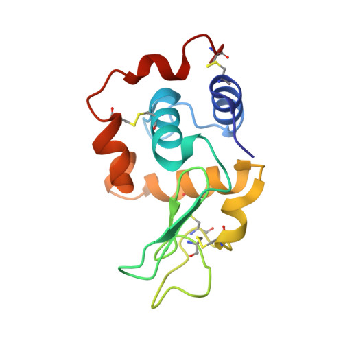
To evaluate the contribution of the amino acid residues on the surface of a protein to its stability, a series of hydrophobic mutant human lysozymes (Val to Gly, Ala, Leu, Ile, Met, and Phe) modified at three different positions on the surface, which are located in the alpha-helix (Val 110), the beta-sheet (Val 2), and the loop (Val 74), were constructed. Their thermodynamic parameters of denaturation and crystal structures were examined by calorimetry and by X-ray crystallography at 100 K, respectively. Differences in the denaturation Gibbs energy change between the wild-type and the hydrophobic mutant proteins ranged from 4.6 to -9.6 kJ/mol, 2.7 to -1.5 kJ/mol, and 3.6 to -0.2 kJ/mol at positions 2, 74, and 110, respectively. The identical substitution at different positions and different substitutions at the same position resulted in different degrees of stabilization. Changes in the stability of the mutant proteins could be evaluated by a unique equation considering the conformational changes due to the substitutions [Funahashi et al. (1999) Protein Eng. 12, 841-850]. For this calculation, secondary structural propensities were newly considered. However, some mutant proteins were not adapted to the equation. The hydration structures around the mutation sites of the exceptional mutant proteins were affected due to the substitutions. The stability changes in the exceptional mutant proteins could be explained by the formation or destruction of the hydration structures. These results suggest that the hydration structure mediated via hydrogen bonds covering the protein surface plays an important role in the conformational stability of the protein.
- Institute for Protein Research, Osaka University, 3-2 Yamadaoka, Suita, Osaka 565-0871, Japan.
Organizational Affiliation: