1ERX

CRYSTAL STRUCTURE OF NITROPHORIN 4 COMPLEXED WITH NO


Experimental Data Snapshot

  • Method: X-RAY DIFFRACTION
  • Resolution: 1.40 Å
  • R-Value Free: 0.240 
  • R-Value Work: 0.170 
  • R-Value Observed: 0.170 

wwPDB Validation   3D Report Full Report


This is version 1.5 of the entry. See complete history


Literature

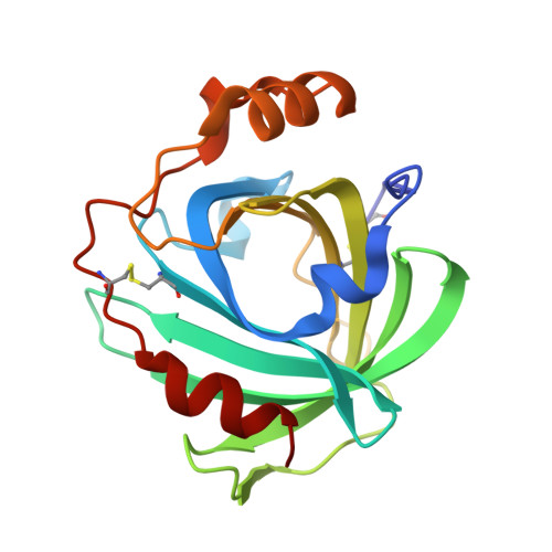
Nitric oxide binding to nitrophorin 4 induces complete distal pocket burial.

Weichsel, A.Andersen, J.F.Roberts, S.A.Montfort, W.R.

(2000) Nat Struct Biol 7: 551-554

  • DOI: https://doi.org/10.1038/76769
  • Primary Citation of Related Structures:  
    1D3S, 1EQD, 1ERX

  • PubMed Abstract: 

    The nitrophorins comprise an unusual family of proteins that use ferric (Fe(III)) heme to transport highly reactive nitric oxide (NO) from the salivary gland of a blood sucking bug to the victim, resulting in vasodilation and reduced blood coagulation. We have determined structures of nitrophorin 4 in complexes with H2O, cyanide and nitric oxide. These structures reveal a remarkable feature: the nitrophorins have a broadly open distal pocket in the absence of NO, but upon NO binding, three or more water molecules are expelled and two loops fold into the distal pocket, resulting in the packing of hydrophobic groups around the NO molecule and increased distortion of the heme. In this way, the protein apparently forms a 'hydrophobic trap' for the NO molecule. The structures are very accurate, ranging between 1.6 and 1.4 A resolutions.


  • Organizational Affiliation

    Department of Biochemistry, University of Arizona, Tucson, Arizona 85721, USA.


Macromolecules
Find similar proteins by:  (by identity cutoff)  |  3D Structure
Entity ID: 1
MoleculeChains Sequence LengthOrganismDetailsImage
NITROPHORIN 4184Rhodnius prolixusMutation(s): 0 
UniProt
Find proteins for Q94734 (Rhodnius prolixus)
Explore Q94734 
Go to UniProtKB:  Q94734
Entity Groups  
Sequence Clusters30% Identity50% Identity70% Identity90% Identity95% Identity100% Identity
UniProt GroupQ94734
Sequence Annotations
Expand
  • Reference Sequence
Small Molecules
Ligands 3 Unique
IDChains Name / Formula / InChI Key2D Diagram3D Interactions
HEV
Query on HEV

Download Ideal Coordinates CCD File 
B [auth A]5,8-DIMETHYL-1,2,3,4-TETRAVINYLPORPHINE-6,7-DIPROPIONIC ACID FERROUS COMPLEX
C36 H32 Fe N4 O4
KPGBVOKZHGYTDW-ZSQORRHCSA-L
CIT
Query on CIT

Download Ideal Coordinates CCD File 
D [auth A]CITRIC ACID
C6 H8 O7
KRKNYBCHXYNGOX-UHFFFAOYSA-N
NO
Query on NO

Download Ideal Coordinates CCD File 
C [auth A]NITRIC OXIDE
N O
ODUCDPQEXGNKDN-UHFFFAOYSA-N
Experimental Data & Validation

Experimental Data

  • Method: X-RAY DIFFRACTION
  • Resolution: 1.40 Å
  • R-Value Free: 0.240 
  • R-Value Work: 0.170 
  • R-Value Observed: 0.170 
  • Space Group: C 1 2 1
Unit Cell:
Length ( Å )Angle ( ˚ )
a = 70.319α = 90
b = 42.636β = 94.19
c = 52.573γ = 90
Software Package:
Software NamePurpose
MADNESSdata collection
SCALAdata scaling
X-PLORmodel building
SHELXL-97refinement
MADNESSdata reduction
CCP4data scaling
X-PLORphasing

Structure Validation

View Full Validation Report


Entry History 

Deposition Data

Revision History  (Full details and data files)

  • Version 1.0: 2000-05-03
    Type: Initial release
  • Version 1.1: 2008-04-27
    Changes: Version format compliance
  • Version 1.2: 2011-07-13
    Changes: Version format compliance
  • Version 1.3: 2017-10-04
    Changes: Refinement description
  • Version 1.4: 2018-01-31
    Changes: Database references
  • Version 1.5: 2018-04-18
    Changes: Data collection