Structure of the VHS domain of human Tom1 (target of myb 1): insights into interactions with proteins and membranes
Misra, S., Beach, B., Hurley, J.H.(2000) Biochemistry 39: 11282-11290
- PubMed: 10985773 Search on PubMed
- DOI: https://doi.org/10.1021/bi0013546
- Primary Citation Related Structures:
1ELK - PubMed Abstract:
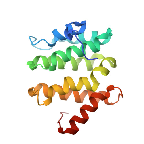
VHS domains are found at the N-termini of select proteins involved in intracellular membrane trafficking. We have determined the crystal structure of the VHS domain of the human Tom1 (target of myb 1) protein to 1.5 A resolution. The domain consists of eight helices arranged in a superhelix. The surface of the domain has two main features: (1) a basic patch on one side due to several conserved positively charged residues on helix 3 and (2) a negatively charged ridge on the opposite side, formed by residues on helix 2. We compare our structure to the recently obtained structure of tandem VHS-FYVE domains from Hrs [Mao, Y., Nickitenko, A., Duan, X., Lloyd, T. E., Wu, M. N., Bellen, H., and Quiocho, F. A. (2000) Cell 100, 447-456]. Key features of the interaction surface between the FYVE and VHS domains of Hrs, involving helices 2 and 4 of the VHS domain, are conserved in the VHS domain of Tom1, even though Tom1 does not have a FYVE domain. We also compare the structures of the VHS domains of Tom1 and Hrs to the recently obtained structure of the ENTH domain of epsin-1 [Hyman, J., Chen, H., Di Fiore, P. P., De Camilli, P., and Brünger, A. T. (2000) J. Cell Biol. 149, 537-546]. Comparison of the two VHS domains and the ENTH domain reveals a conserved surface, composed of helices 2 and 4, that is utilized for protein-protein interactions. In addition, VHS domain-containing proteins are often localized to membranes. We suggest that the conserved positively charged surface of helix 3 in VHS and ENTH domains plays a role in membrane binding.
- Laboratory of Molecular Biology, National Institute of Diabetes and Digestive and Kidney Diseases, National Institutes of Health, Bethesda, Maryland 20892-0580, USA.
Organizational Affiliation: