1EKI

AVERAGE SOLUTION STRUCTURE OF D(TTGGCCAA)2 BOUND TO CHROMOMYCIN-A3 AND COBALT

  • Classification: DNA
  • Mutation(s): No 

  • Deposited: 2000-03-08 Released: 2000-03-20 
  • Deposition Author(s): Gochin, M.

Experimental Data Snapshot

  • Method: SOLUTION NMR
  • Conformers Calculated: 
  • Conformers Submitted: 
  • Selection Criteria: average structure 

wwPDB Validation   3D Report Full Report


This is version 2.1 of the entry. See complete history


Literature

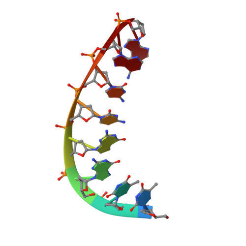
A high-resolution structure of a DNA-chromomycin-Co(II) complex determined from pseudocontact shifts in nuclear magnetic resonance.

Gochin, M.

(2000) Structure 8: 441-452

  • DOI: https://doi.org/10.1016/s0969-2126(00)00124-6
  • Primary Citation of Related Structures:  
    1EKH, 1EKI

  • PubMed Abstract: 

    The drug chromomycin-A(3) binds to the minor groove of DNA and requires a divalent metal ion for complex formation. (1)H, (31)P and (13)C pseudocontact shifts occurring in the presence of a tightly bound divalent cobalt ion in the complex between d(TTGGCCAA)(2) and chromomycin-A(3) have been used to determine the structure of the complex. The accuracy of the structure was verified by validation with nuclear Overhauser enhancements (NOEs) and J-coupling constants not used in the structure calculation. The final structure was determined to 0.7 A resolution. The structure was compared with a structure obtained in an earlier study using NOEs, in order to assess the accuracy of NOEs in giving global structural information for a DNA complex. Although some basic features of the structures agreed, they differed substantially in the fine structural details and in the DNA axis curvature generated by the drug. The distortion of base-pair planarity that was observed in the NOE structure was not seen in our structure. Differences in drug orientation and hydrogen bonding also occurred. The curvature and elongation of the DNA that was obtained previously was not found to occur in our study. The use of pseudocontact shifts has enabled us to obtain a high-precision global structure of the chromomycin-DNA complex, which provides an accurate template on which to consider targeting minor groove binding drugs. The effect of such binding is not propagated far along the helix but is restricted to a local kink in the axis that reverts to its original direction within four base pairs.


  • Organizational Affiliation

    Department of Microbiology, University of the Pacific School of Dentistry, San Francisco, CA 94115, USA. miriam@picasso. ucsf.edu


Macromolecules

Find similar nucleic acids by:  Sequence   |   3D Structure  

Entity ID: 1
MoleculeChains LengthOrganismImage
DNA (5'-D(*TP*TP*GP*GP*CP*CP*AP*A)-3')
A, B
8N/A
Sequence Annotations
Expand
  • Reference Sequence
Oligosaccharides

Help

Entity ID: 2
MoleculeChains Length2D Diagram Glycosylation3D Interactions
2,6-dideoxy-4-O-methyl-alpha-D-galactopyranose-(1-3)-4-O-acetyl-2,6-dideoxy-beta-D-galactopyranose
C, D
2N/A
Glycosylation Resources
GlyTouCan:  G63753LU
GlyCosmos:  G63753LU
Entity ID: 3
MoleculeChains Length2D Diagram Glycosylation3D Interactions
3-C-methyl-4-O-acetyl-alpha-L-Olivopyranose-(1-3)-beta-D-Olivopyranose-(1-3)-beta-D-Olivopyranose
E, F
3N/A
Glycosylation Resources
GlyTouCan:  G79611HT
GlyCosmos:  G79611HT
Small Molecules
Ligands 2 Unique
IDChains Name / Formula / InChI Key2D Diagram3D Interactions
CPH
Query on CPH

Download Ideal Coordinates CCD File 
H [auth A],
I [auth B]
(1S)-5-deoxy-1-O-methyl-1-C-[(2R,3S)-3,5,7,10-tetrahydroxy-6-methyl-4-oxo-1,2,3,4-tetrahydroanthracen-2-yl]-D-xylulose
C21 H24 O9
AOCJXLJIUYHXRS-PKRZCTMMSA-N
CO
Query on CO

Download Ideal Coordinates CCD File 
G [auth A]COBALT (II) ION
Co
XLJKHNWPARRRJB-UHFFFAOYSA-N
Experimental Data & Validation

Experimental Data

  • Method: SOLUTION NMR
  • Conformers Calculated: 
  • Conformers Submitted: 
  • Selection Criteria: average structure 

Structure Validation

View Full Validation Report


Entry History 

Deposition Data

  • Released Date: 2000-03-20 
  • Deposition Author(s): Gochin, M.

Revision History  (Full details and data files)

  • Version 1.0: 2000-03-20
    Type: Initial release
  • Version 1.1: 2008-04-27
    Changes: Version format compliance
  • Version 1.2: 2011-07-13
    Changes: Version format compliance
  • Version 1.3: 2011-12-28
    Changes: Advisory
  • Version 2.0: 2020-07-29
    Type: Remediation
    Reason: Carbohydrate remediation
    Changes: Advisory, Atomic model, Data collection, Derived calculations, Structure summary
  • Version 2.1: 2024-05-01
    Changes: Data collection, Database references, Structure summary