6ZEK

Crystal structure of mouse CSAD


Experimental Data Snapshot

  • Method: X-RAY DIFFRACTION
  • Resolution: 2.10 Å
  • R-Value Free: 0.235 
  • R-Value Work: 0.179 
  • R-Value Observed: 0.181 

wwPDB Validation   3D Report Full Report


This is version 1.1 of the entry. See complete history


Literature

Structure and substrate specificity determinants of the taurine biosynthetic enzyme cysteine sulphinic acid decarboxylase.

Mahootchi, E.Raasakka, A.Luan, W.Muruganandam, G.Loris, R.Haavik, J.Kursula, P.

(2021) J Struct Biol 213: 107674-107674

  • DOI: https://doi.org/10.1016/j.jsb.2020.107674
  • Primary Citation of Related Structures:  
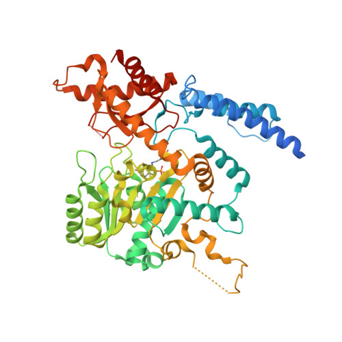
    6ZEK

  • PubMed Abstract: 

    Pyridoxal 5́-phosphate (PLP) is an important cofactor for amino acid decarboxylases with many biological functions, including the synthesis of signalling molecules, such as serotonin, dopamine, histamine, γ-aminobutyric acid, and taurine. Taurine is an abundant amino acid with multiple physiological functions, including osmoregulation, pH regulation, antioxidative protection, and neuromodulation. In mammalian tissues, taurine is mainly produced by decarboxylation of cysteine sulphinic acid to hypotaurine, catalysed by the PLP-dependent cysteine sulphinic acid decarboxylase (CSAD), followed by oxidation of the product to taurine. We determined the crystal structure of mouse CSAD and compared it to other PLP-dependent decarboxylases in order to identify determinants of substrate specificity and catalytic activity. Recognition of the substrate involves distinct side chains forming the substrate-binding cavity. In addition, the backbone conformation of a buried active-site loop appears to be a critical determinant for substrate side chain binding in PLP-dependent decarboxylases. Phe94 was predicted to affect substrate specificity, and its mutation to serine altered both the catalytic properties of CSAD and its stability. Using small-angle X-ray scattering, we further showed that CSAD presents open/close motions in solution. The structure of apo-CSAD indicates that the active site gets more ordered upon internal aldimine formation. Taken together, the results highlight details of substrate recognition in PLP-dependent decarboxylases and provide starting points for structure-based inhibitor design with the aim of affecting the biosynthesis of taurine and other abundant amino acid metabolites.


  • Organizational Affiliation

    Department of Biomedicine, University of Bergen, Bergen, Norway.


Macromolecules
Find similar proteins by:  (by identity cutoff)  |  3D Structure
Entity ID: 1
MoleculeChains Sequence LengthOrganismDetailsImage
Cysteine sulfinic acid decarboxylase
A, B, C, D
521Mus musculusMutation(s): 0 
Gene Names: Csad
EC: 4.1.1.29 (PDB Primary Data), 4.1.1.11 (PDB Primary Data)
UniProt
Find proteins for Q9DBE0 (Mus musculus)
Explore Q9DBE0 
Go to UniProtKB:  Q9DBE0
Entity Groups  
Sequence Clusters30% Identity50% Identity70% Identity90% Identity95% Identity100% Identity
UniProt GroupQ9DBE0
Sequence Annotations
Expand
  • Reference Sequence
Small Molecules
Ligands 4 Unique
IDChains Name / Formula / InChI Key2D Diagram3D Interactions
SO4
Query on SO4

Download Ideal Coordinates CCD File 
E [auth A]SULFATE ION
O4 S
QAOWNCQODCNURD-UHFFFAOYSA-L
GOL
Query on GOL

Download Ideal Coordinates CCD File 
J [auth B],
P [auth C],
Q [auth C],
V [auth D]
GLYCEROL
C3 H8 O3
PEDCQBHIVMGVHV-UHFFFAOYSA-N
CO
Query on CO

Download Ideal Coordinates CCD File 
F [auth A],
K [auth B],
R [auth C],
W [auth D]
COBALT (II) ION
Co
XLJKHNWPARRRJB-UHFFFAOYSA-N
CL
Query on CL

Download Ideal Coordinates CCD File 
G [auth A]
H [auth A]
I [auth A]
L [auth B]
M [auth B]
G [auth A],
H [auth A],
I [auth A],
L [auth B],
M [auth B],
N [auth B],
O [auth B],
S [auth C],
T [auth C],
U [auth C],
X [auth D],
Y [auth D],
Z [auth D]
CHLORIDE ION
Cl
VEXZGXHMUGYJMC-UHFFFAOYSA-M
Modified Residues  1 Unique
IDChains TypeFormula2D DiagramParent
LLP
Query on LLP
A, B, C, D
L-PEPTIDE LINKINGC14 H22 N3 O7 PLYS
Experimental Data & Validation

Experimental Data

  • Method: X-RAY DIFFRACTION
  • Resolution: 2.10 Å
  • R-Value Free: 0.235 
  • R-Value Work: 0.179 
  • R-Value Observed: 0.181 
  • Space Group: P 1 21 1
Unit Cell:
Length ( Å )Angle ( ˚ )
a = 72.94α = 90
b = 113.26β = 95.808
c = 113.37γ = 90
Software Package:
Software NamePurpose
PHASERphasing
PHENIXrefinement
XDSdata reduction
XSCALEdata scaling

Structure Validation

View Full Validation Report


Entry History 

Deposition Data

Revision History  (Full details and data files)

  • Version 1.0: 2021-04-28
    Type: Initial release
  • Version 1.1: 2024-01-24
    Changes: Advisory, Data collection, Database references, Refinement description