6W2Y

CryoEM Structure of GABAB1b Homodimer


Experimental Data Snapshot

  • Method: ELECTRON MICROSCOPY
  • Resolution: 3.20 Å
  • Aggregation State: PARTICLE 
  • Reconstruction Method: SINGLE PARTICLE 

wwPDB Validation   3D Report Full Report


This is version 2.1 of the entry. See complete history


Literature

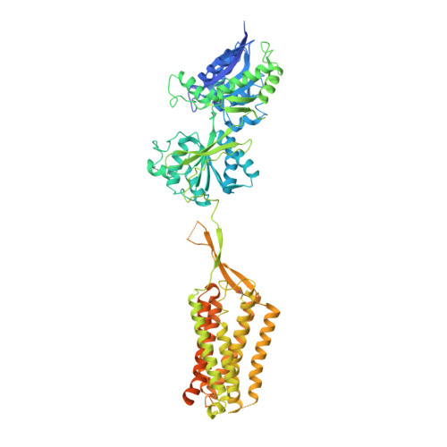
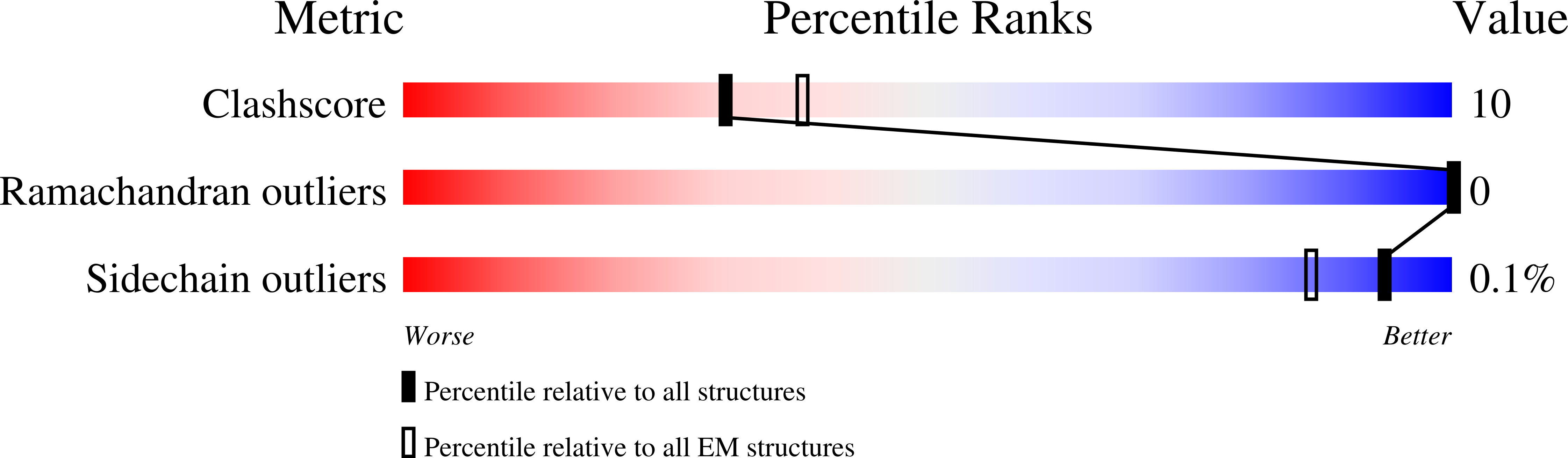
Structures of metabotropic GABABreceptor.

Papasergi-Scott, M.M.Robertson, M.J.Seven, A.B.Panova, O.Mathiesen, J.M.Skiniotis, G.

(2020) Nature 584: 310-314

  • DOI: https://doi.org/10.1038/s41586-020-2469-4
  • Primary Citation of Related Structures:  
    6W2X, 6W2Y

  • PubMed Abstract: 

    Stimulation of the metabotropic GABA B receptor by γ-aminobutyric acid (GABA) results in prolonged inhibition of neurotransmission, which is central to brain physiology 1 . GABA B belongs to family C of the G-protein-coupled receptors, which operate as dimers to transform synaptic neurotransmitter signals into a cellular response through the binding and activation of heterotrimeric G proteins 2,3 . However, GABA B is unique in its function as an obligate heterodimer in which agonist binding and G-protein activation take place on distinct subunits 4,5 . Here we present cryo-electron microscopy structures of heterodimeric and homodimeric full-length GABA B receptors. Complemented by cellular signalling assays and atomistic simulations, these structures reveal that extracellular loop 2 (ECL2) of GABA B has an essential role in relaying structural transitions by ordering the linker that connects the extracellular ligand-binding domain to the transmembrane region. Furthermore, the ECL2 of each of the subunits of GABA B caps and interacts with the hydrophilic head of a phospholipid that occupies the extracellular half of the transmembrane domain, thereby providing a potentially crucial link between ligand binding and the receptor core that engages G proteins. These results provide a starting framework through which to decipher the mechanistic modes of signal transduction mediated by GABA B dimers, and have important implications for rational drug design that targets these receptors.


  • Organizational Affiliation

    Department of Molecular and Cellular Physiology, Stanford University School of Medicine, Stanford, CA, USA.


Macromolecules
Find similar proteins by:  (by identity cutoff)  |  3D Structure
Entity ID: 1
MoleculeChains Sequence LengthOrganismDetailsImage
Gamma-aminobutyric acid type B receptor subunit 1
A, B
829Homo sapiensMutation(s): 0 
Gene Names: GABBR1GPRC3A
Membrane Entity: Yes 
UniProt & NIH Common Fund Data Resources
Find proteins for Q9UBS5 (Homo sapiens)
Explore Q9UBS5 
Go to UniProtKB:  Q9UBS5
PHAROS:  Q9UBS5
GTEx:  ENSG00000204681 
Entity Groups  
Sequence Clusters30% Identity50% Identity70% Identity90% Identity95% Identity100% Identity
UniProt GroupQ9UBS5
Sequence Annotations
Expand
  • Reference Sequence
Oligosaccharides

Help

Entity ID: 2
MoleculeChains Length2D Diagram Glycosylation3D Interactions
2-acetamido-2-deoxy-beta-D-glucopyranose-(1-4)-2-acetamido-2-deoxy-beta-D-glucopyranose
C, D
2N-Glycosylation
Glycosylation Resources
GlyTouCan:  G42666HT
GlyCosmos:  G42666HT
GlyGen:  G42666HT
Small Molecules
Ligands 4 Unique
IDChains Name / Formula / InChI Key2D Diagram3D Interactions
L9Q
Query on L9Q

Download Ideal Coordinates CCD File 
H [auth A],
M [auth B]
(1S)-2-{[(S)-(2-aminoethoxy)(hydroxy)phosphoryl]oxy}-1-[(octadecanoyloxy)methyl]ethyl (9Z)-octadec-9-enoate
C41 H80 N O8 P
JQKOHRZNEOQNJE-VYCVAHKRSA-N
SGG
Query on SGG

Download Ideal Coordinates CCD File 
I [auth A],
N [auth B]
[(2~{S})-3-[[(1~{S})-1-(3,4-dichlorophenyl)ethyl]amino]-2-oxidanyl-propyl]-(phenylmethyl)phosphinic acid
C18 H22 Cl2 N O3 P
ZODSPDOOCZZEIM-BBRMVZONSA-N
NAG
Query on NAG

Download Ideal Coordinates CCD File 
E [auth A],
F [auth A],
J [auth B],
K [auth B]
2-acetamido-2-deoxy-beta-D-glucopyranose
C8 H15 N O6
OVRNDRQMDRJTHS-FMDGEEDCSA-N
MG
Query on MG

Download Ideal Coordinates CCD File 
G [auth A],
L [auth B]
MAGNESIUM ION
Mg
JLVVSXFLKOJNIY-UHFFFAOYSA-N
Experimental Data & Validation

Experimental Data

  • Method: ELECTRON MICROSCOPY
  • Resolution: 3.20 Å
  • Aggregation State: PARTICLE 
  • Reconstruction Method: SINGLE PARTICLE 
EM Software:
TaskSoftware PackageVersion
RECONSTRUCTIONRELION3.0

Structure Validation

View Full Validation Report


Entry History & Funding Information

Deposition Data


Funding OrganizationLocationGrant Number
National Institutes of Health/National Institute of Neurological Disorders and Stroke (NIH/NINDS)United States1R01NS092695-01

Revision History  (Full details and data files)

  • Version 1.0: 2020-07-01
    Type: Initial release
  • Version 1.1: 2020-07-08
    Changes: Database references
  • Version 2.0: 2020-07-29
    Type: Remediation
    Reason: Carbohydrate remediation
    Changes: Advisory, Atomic model, Data collection, Derived calculations, Structure summary
  • Version 2.1: 2020-08-26
    Changes: Database references, Structure summary