6PZ8

MERS S0 trimer in complex with variable domain of antibody G2


Experimental Data Snapshot

  • Method: ELECTRON MICROSCOPY
  • Resolution: 4.19 Å
  • Aggregation State: PARTICLE 
  • Reconstruction Method: SINGLE PARTICLE 

wwPDB Validation   3D Report Full Report


This is version 2.0 of the entry. See complete history


Literature

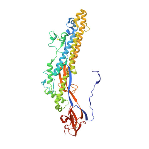
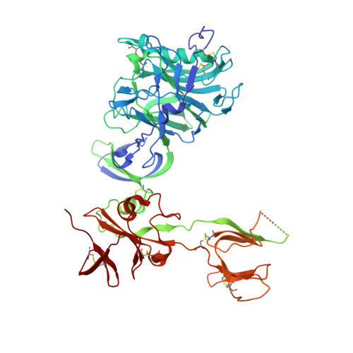
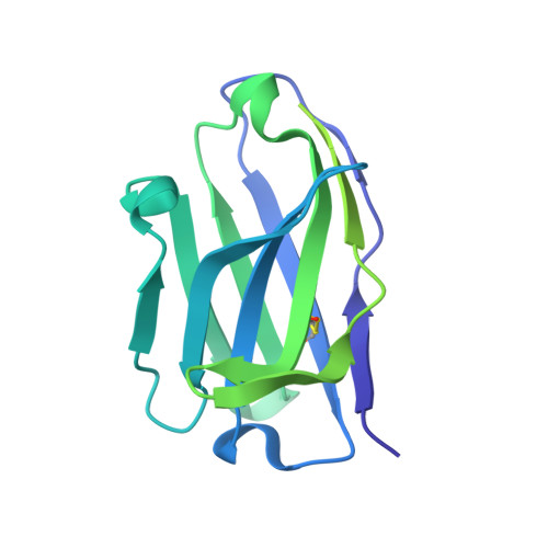
Structural Definition of a Neutralization-Sensitive Epitope on the MERS-CoV S1-NTD.

Wang, N.Rosen, O.Wang, L.Turner, H.L.Stevens, L.J.Corbett, K.S.Bowman, C.A.Pallesen, J.Shi, W.Zhang, Y.Leung, K.Kirchdoerfer, R.N.Becker, M.M.Denison, M.R.Chappell, J.D.Ward, A.B.Graham, B.S.McLellan, J.S.

(2019) Cell Rep 28: 3395-3405.e6

  • DOI: https://doi.org/10.1016/j.celrep.2019.08.052
  • Primary Citation of Related Structures:  
    6PXG, 6PXH, 6PZ8

  • PubMed Abstract: 

    Middle East respiratory syndrome coronavirus (MERS-CoV) emerged into the human population in 2012 and has caused substantial morbidity and mortality. Potently neutralizing antibodies targeting the receptor-binding domain (RBD) on MERS-CoV spike (S) protein have been characterized, but much less is known about antibodies targeting non-RBD epitopes. Here, we report the structural and functional characterization of G2, a neutralizing antibody targeting the MERS-CoV S1 N-terminal domain (S1-NTD). Structures of G2 alone and in complex with the MERS-CoV S1-NTD define a site of vulnerability comprising two loops, each of which contain a residue mutated in G2-escape variants. Cell-surface binding studies and in vitro competition experiments demonstrate that G2 strongly disrupts the attachment of MERS-CoV S to its receptor, dipeptidyl peptidase-4 (DPP4), with the inhibition requiring the native trimeric S conformation. These results advance our understanding of antibody-mediated neutralization of coronaviruses and should facilitate the development of immunotherapeutics and vaccines against MERS-CoV.


  • Organizational Affiliation

    Department of Molecular Biosciences, The University of Texas at Austin, Austin, TX 78712, USA.


Macromolecules
Find similar proteins by:  (by identity cutoff)  |  3D Structure
Entity ID: 1
MoleculeChains Sequence LengthOrganismDetailsImage
S protein
A, E, I
472Middle East respiratory syndrome-related coronavirusMutation(s): 0 
UniProt
Find proteins for K9N5Q8 (Middle East respiratory syndrome-related coronavirus (isolate United Kingdom/H123990006/2012))
Explore K9N5Q8 
Go to UniProtKB:  K9N5Q8
Entity Groups  
Sequence Clusters30% Identity50% Identity70% Identity90% Identity95% Identity100% Identity
UniProt GroupK9N5Q8
Sequence Annotations
Expand
  • Reference Sequence
Find similar proteins by:  (by identity cutoff)  |  3D Structure
Entity ID: 2
MoleculeChains Sequence LengthOrganismDetailsImage
S proteinB,
F [auth J],
J [auth F]
726Middle East respiratory syndrome-related coronavirusMutation(s): 0 
UniProt
Find proteins for K9N5Q8 (Middle East respiratory syndrome-related coronavirus (isolate United Kingdom/H123990006/2012))
Explore K9N5Q8 
Go to UniProtKB:  K9N5Q8
Entity Groups  
Sequence Clusters30% Identity50% Identity70% Identity90% Identity95% Identity100% Identity
UniProt GroupK9N5Q8
Sequence Annotations
Expand
  • Reference Sequence
Find similar proteins by:  (by identity cutoff)  |  3D Structure
Entity ID: 3
MoleculeChains Sequence LengthOrganismDetailsImage
G2 heavy chainC [auth H],
G [auth C],
K [auth D]
229Mus musculusMutation(s): 0 
Entity Groups  
Sequence Clusters30% Identity50% Identity70% Identity90% Identity95% Identity100% Identity
Sequence Annotations
Expand
  • Reference Sequence
Find similar proteins by:  (by identity cutoff)  |  3D Structure
Entity ID: 4
MoleculeChains Sequence LengthOrganismDetailsImage
G2 light chainD [auth L],
H [auth G],
L [auth K]
218Mus musculusMutation(s): 0 
Entity Groups  
Sequence Clusters30% Identity50% Identity70% Identity90% Identity95% Identity100% Identity
Sequence Annotations
Expand
  • Reference Sequence
Oligosaccharides

Help

Entity ID: 6
MoleculeChains Length2D Diagram Glycosylation3D Interactions
beta-D-mannopyranose-(1-4)-2-acetamido-2-deoxy-beta-D-glucopyranose-(1-4)-2-acetamido-2-deoxy-beta-D-glucopyranoseGA [auth g],
Q,
Y
3N-Glycosylation
Glycosylation Resources
GlyTouCan:  G15407YE
GlyCosmos:  G15407YE
GlyGen:  G15407YE
Entity ID: 7
MoleculeChains Length2D Diagram Glycosylation3D Interactions
alpha-D-mannopyranose-(1-3)-[alpha-D-mannopyranose-(1-6)]beta-D-mannopyranose-(1-4)-2-acetamido-2-deoxy-beta-D-glucopyranose-(1-4)-2-acetamido-2-deoxy-beta-D-glucopyranoseAA [auth a],
IA [auth i],
S
5N-Glycosylation
Glycosylation Resources
GlyTouCan:  G22768VO
GlyCosmos:  G22768VO
GlyGen:  G22768VO
Small Molecules
Ligands 1 Unique
IDChains Name / Formula / InChI Key2D Diagram3D Interactions
NAG
Query on NAG

Download Ideal Coordinates CCD File 
AB [auth F]
BB [auth F]
KA [auth A]
LA [auth B]
MA [auth B]
AB [auth F],
BB [auth F],
KA [auth A],
LA [auth B],
MA [auth B],
NA [auth B],
OA [auth B],
PA [auth B],
QA [auth E],
RA [auth J],
SA [auth J],
TA [auth J],
UA [auth J],
VA [auth J],
WA [auth I],
XA [auth F],
YA [auth F],
ZA [auth F]
2-acetamido-2-deoxy-beta-D-glucopyranose
C8 H15 N O6
OVRNDRQMDRJTHS-FMDGEEDCSA-N
Experimental Data & Validation

Experimental Data

  • Method: ELECTRON MICROSCOPY
  • Resolution: 4.19 Å
  • Aggregation State: PARTICLE 
  • Reconstruction Method: SINGLE PARTICLE 
EM Software:
TaskSoftware PackageVersion
RECONSTRUCTIONcryoSPARC2.5

Structure Validation

View Full Validation Report


Entry History & Funding Information

Deposition Data


Funding OrganizationLocationGrant Number
National Institutes of Health/National Institute Of Allergy and Infectious Diseases (NIH/NIAID)United StatesAI127521

Revision History  (Full details and data files)

  • Version 1.0: 2019-10-09
    Type: Initial release
  • Version 1.1: 2019-12-18
    Changes: Author supporting evidence
  • Version 2.0: 2020-07-29
    Type: Remediation
    Reason: Carbohydrate remediation
    Changes: Atomic model, Data collection, Derived calculations, Structure summary