6HN4

Leucine-zippered human insulin receptor ectodomain with single bound insulin - "lower" membrane-proximal part


Experimental Data Snapshot

  • Method: ELECTRON MICROSCOPY
  • Resolution: 4.20 Å
  • Aggregation State: PARTICLE 
  • Reconstruction Method: SINGLE PARTICLE 

wwPDB Validation   3D Report Full Report


This is version 1.4 of the entry. See complete history


Literature

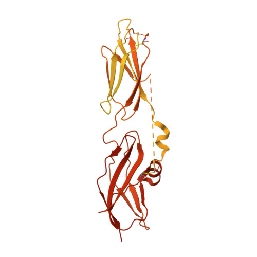
The signalling conformation of the insulin receptor ectodomain.

Weis, F.Menting, J.G.Margetts, M.B.Chan, S.J.Xu, Y.Tennagels, N.Wohlfart, P.Langer, T.Muller, C.W.Dreyer, M.K.Lawrence, M.C.

(2018) Nat Commun 9: 4420-4420

  • DOI: https://doi.org/10.1038/s41467-018-06826-6
  • Primary Citation of Related Structures:  
    6HN4, 6HN5

  • PubMed Abstract: 

    Understanding the structural biology of the insulin receptor and how it signals is of key importance in the development of insulin analogs to treat diabetes. We report here a cryo-electron microscopy structure of a single insulin bound to a physiologically relevant, high-affinity version of the receptor ectodomain, the latter generated through attachment of C-terminal leucine zipper elements to overcome the conformational flexibility associated with ectodomain truncation. The resolution of the cryo-electron microscopy maps is 3.2 Å in the insulin-binding region and 4.2 Å in the membrane-proximal region. The structure reveals how the membrane proximal domains of the receptor come together to effect signalling and how insulin's negative cooperativity of binding likely arises. Our structure further provides insight into the high affinity of certain super-mitogenic insulins. Together, these findings provide a new platform for insulin analog investigation and design.


  • Organizational Affiliation

    European Molecular Biology Laboratory (EMBL), Structural and Computational Biology Unit, Meyerhofstraße 1, 69117, Heidelberg, Germany.


Macromolecules
Find similar proteins by:  (by identity cutoff)  |  3D Structure
Entity ID: 1
MoleculeChains Sequence LengthOrganismDetailsImage
Insulin receptor,Insulin receptor,General control protein GCN4A [auth E],
B [auth F]
930Homo sapiensSaccharomyces cerevisiae S288CMutation(s): 7 
Gene Names: INSRGCN4AAS3ARG9YEL009C
EC: 2.7.10.1
UniProt & NIH Common Fund Data Resources
Find proteins for P03069 (Saccharomyces cerevisiae (strain ATCC 204508 / S288c))
Explore P03069 
Go to UniProtKB:  P03069
Find proteins for P06213 (Homo sapiens)
Explore P06213 
Go to UniProtKB:  P06213
PHAROS:  P06213
GTEx:  ENSG00000171105 
Entity Groups  
Sequence Clusters30% Identity50% Identity70% Identity90% Identity95% Identity100% Identity
UniProt GroupsP06213P03069
Sequence Annotations
Expand
  • Reference Sequence
Experimental Data & Validation

Experimental Data

  • Method: ELECTRON MICROSCOPY
  • Resolution: 4.20 Å
  • Aggregation State: PARTICLE 
  • Reconstruction Method: SINGLE PARTICLE 
EM Software:
TaskSoftware PackageVersion
MODEL REFINEMENTPHENIX1.13-2998-1692
RECONSTRUCTIONRELION2.1

Structure Validation

View Full Validation Report


Entry History & Funding Information

Deposition Data


Funding OrganizationLocationGrant Number
National Health and Medical Research Council (Australia)AustraliaAPP1099595
National Health and Medical Research Council (Australia)AustraliaAPP1128553
National Institutes of Health/National Institute of Diabetes and Digestive and Kidney Disease (NIH/NIDDK)United StatesDK013914
National Institutes of Health/National Institute of Diabetes and Digestive and Kidney Disease (NIH/NIDDK)United StatesUC DRTC DK020595

Revision History  (Full details and data files)

  • Version 1.0: 2018-11-21
    Type: Initial release
  • Version 1.1: 2019-11-06
    Changes: Data collection, Refinement description
  • Version 1.2: 2019-12-18
    Changes: Other
  • Version 1.3: 2020-07-29
    Type: Remediation
    Reason: Carbohydrate remediation
    Changes: Data collection, Derived calculations, Structure summary
  • Version 1.4: 2022-03-30
    Changes: Author supporting evidence, Database references, Structure summary