5W76

Crystal Structure of Reconstructed Bacterial Elongation Factor Node 168


Experimental Data Snapshot

  • Method: X-RAY DIFFRACTION
  • Resolution: 2.15 Å
  • R-Value Free: 0.223 
  • R-Value Work: 0.190 
  • R-Value Observed: 0.192 

wwPDB Validation   3D Report Full Report


Ligand Structure Quality Assessment 


This is version 1.1 of the entry. See complete history


Literature

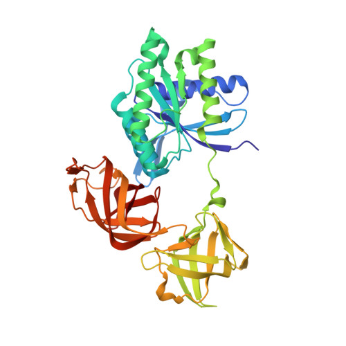
Structural and Dynamics Comparison of Thermostability in Ancient, Modern, and Consensus Elongation Factor Tus.

Okafor, C.D.Pathak, M.C.Fagan, C.E.Bauer, N.C.Cole, M.F.Gaucher, E.A.Ortlund, E.A.

(2018) Structure 26: 118-129.e3

  • DOI: https://doi.org/10.1016/j.str.2017.11.018
  • Primary Citation of Related Structures:  
    5W75, 5W76, 5W7Q

  • PubMed Abstract: 

    Rationally engineering thermostability in proteins would create enzymes and receptors that function under harsh industrial applications. Several sequence-based approaches can generate thermostable variants of mesophilic proteins. To gain insight into the mechanisms by which proteins become more stable, we use structural and dynamic analyses to compare two popular approaches, ancestral sequence reconstruction (ASR) and the consensus method, used to generate thermostable variants of Elongation Factor Thermo-unstable (EF-Tu). We present crystal structures of ancestral and consensus EF-Tus, accompanied by molecular dynamics simulations aimed at probing the strategies employed to enhance thermostability. All proteins adopt crystal structures similar to extant EF-Tus, revealing no difference in average structure between the methods. Molecular dynamics reveals that ASR-generated sequences retain dynamic properties similar to extant, thermostable EF-Tu from Thermus aquaticus, while consensus EF-Tu dynamics differ from evolution-based sequences. This work highlights the advantage of ASR for engineering thermostability while preserving natural motions in multidomain proteins.


  • Organizational Affiliation

    Department of Biochemistry, Emory University School of Medicine, Atlanta, GA 30322, USA.


Macromolecules
Find similar proteins by:  (by identity cutoff)  |  3D Structure
Entity ID: 1
MoleculeChains Sequence LengthOrganismDetailsImage
Ancestral Elogation Factor N153385synthetic constructMutation(s): 0 
Entity Groups  
Sequence Clusters30% Identity50% Identity70% Identity90% Identity95% Identity100% Identity
Sequence Annotations
Expand
  • Reference Sequence
Small Molecules
Ligands 5 Unique
IDChains Name / Formula / InChI Key2D Diagram3D Interactions
GDP
Query on GDP

Download Ideal Coordinates CCD File 
E [auth A]GUANOSINE-5'-DIPHOSPHATE
C10 H15 N5 O11 P2
QGWNDRXFNXRZMB-UUOKFMHZSA-N
PGE
Query on PGE

Download Ideal Coordinates CCD File 
D [auth A]TRIETHYLENE GLYCOL
C6 H14 O4
ZIBGPFATKBEMQZ-UHFFFAOYSA-N
PEG
Query on PEG

Download Ideal Coordinates CCD File 
F [auth A],
G [auth A]
DI(HYDROXYETHYL)ETHER
C4 H10 O3
MTHSVFCYNBDYFN-UHFFFAOYSA-N
SO4
Query on SO4

Download Ideal Coordinates CCD File 
C [auth A]SULFATE ION
O4 S
QAOWNCQODCNURD-UHFFFAOYSA-L
MG
Query on MG

Download Ideal Coordinates CCD File 
B [auth A]MAGNESIUM ION
Mg
JLVVSXFLKOJNIY-UHFFFAOYSA-N
Experimental Data & Validation

Experimental Data

  • Method: X-RAY DIFFRACTION
  • Resolution: 2.15 Å
  • R-Value Free: 0.223 
  • R-Value Work: 0.190 
  • R-Value Observed: 0.192 
  • Space Group: P 21 21 21
Unit Cell:
Length ( Å )Angle ( ˚ )
a = 57.326α = 90
b = 73.357β = 90
c = 114.377γ = 90
Software Package:
Software NamePurpose
HKL-2000data scaling
REFMACrefinement
PDB_EXTRACTdata extraction
HKL-2000data reduction
PHASERphasing

Structure Validation

View Full Validation Report


Ligand Structure Quality Assessment 


Entry History 

Deposition Data

Revision History  (Full details and data files)

  • Version 1.0: 2018-04-25
    Type: Initial release
  • Version 1.1: 2024-03-13
    Changes: Data collection, Database references, Derived calculations