5L7I

Structure of human Smoothened in complex with Vismodegib


Experimental Data Snapshot

  • Method: X-RAY DIFFRACTION
  • Resolution: 3.30 Å
  • R-Value Free: 0.257 
  • R-Value Work: 0.245 
  • R-Value Observed: 0.245 

wwPDB Validation   3D Report Full Report


Ligand Structure Quality Assessment 


This is version 1.5 of the entry. See complete history


Literature

Structural basis of Smoothened regulation by its extracellular domains.

Byrne, E.F.Sircar, R.Miller, P.S.Hedger, G.Luchetti, G.Nachtergaele, S.Tully, M.D.Mydock-McGrane, L.Covey, D.F.Rambo, R.P.Sansom, M.S.Newstead, S.Rohatgi, R.Siebold, C.

(2016) Nature 535: 517-522

  • DOI: https://doi.org/10.1038/nature18934
  • Primary Citation of Related Structures:  
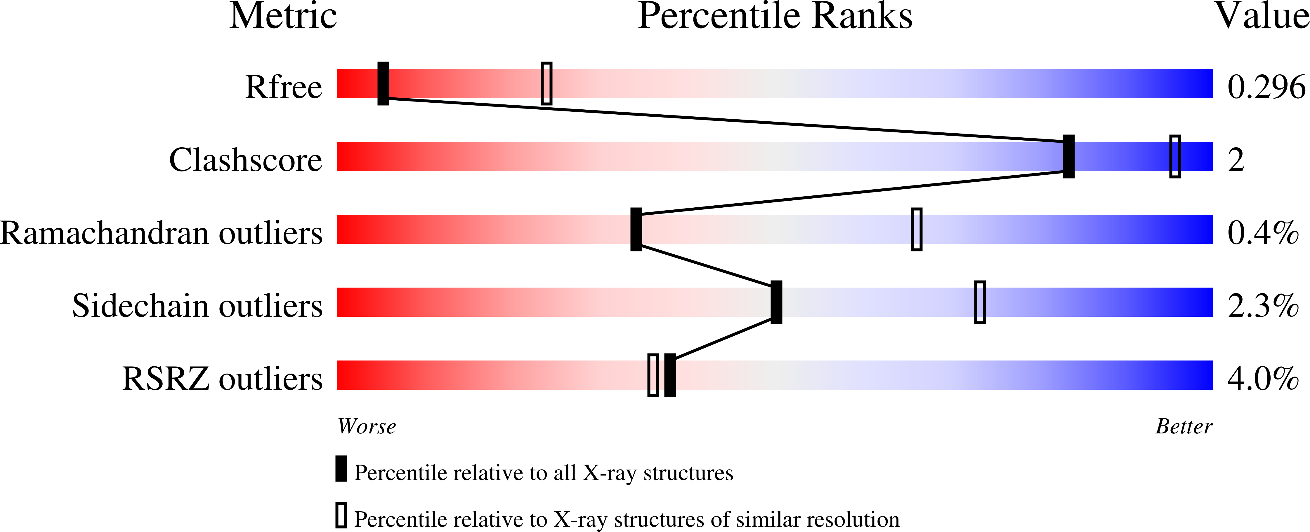
    5L7D, 5L7I

  • PubMed Abstract: 

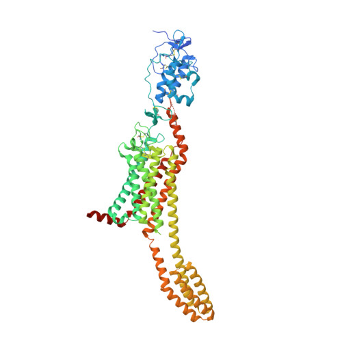
    Developmental signals of the Hedgehog (Hh) and Wnt families are transduced across the membrane by Frizzledclass G-protein-coupled receptors (GPCRs) composed of both a heptahelical transmembrane domain (TMD) and an extracellular cysteine-rich domain (CRD). How the large extracellular domains of GPCRs regulate signalling by the TMD is unknown. We present crystal structures of the Hh signal transducer and oncoprotein Smoothened, a GPCR that contains two distinct ligand-binding sites: one in its TMD and one in the CRD. The CRD is stacked a top the TMD, separated by an intervening wedge-like linker domain. Structure-guided mutations show that the interface between the CRD, linker domain and TMD stabilizes the inactive state of Smoothened. Unexpectedly, we find a cholesterol molecule bound to Smoothened in the CRD binding site. Mutations predicted to prevent cholesterol binding impair the ability of Smoothened to transmit native Hh signals. Binding of a clinically used antagonist, vismodegib, to the TMD induces a conformational change that is propagated to the CRD, resulting in loss of cholesterol from the CRD-linker domain-TMD interface. Our results clarify the structural mechanism by which the activity of a GPCR is controlled by ligand-regulated interactions between its extracellular and transmembrane domains.


  • Organizational Affiliation

    Division of Structural Biology, Wellcome Trust Centre for Human Genetics, University of Oxford, Oxford, UK.


Macromolecules
Find similar proteins by:  (by identity cutoff)  |  3D Structure
Entity ID: 1
MoleculeChains Sequence LengthOrganismDetailsImage
Smoothened homolog,Soluble cytochrome b562,Smoothened homolog
A, B
638Homo sapiensEscherichia coliMutation(s): 0 
Gene Names: SMOSMOHcybC
Membrane Entity: Yes 
UniProt & NIH Common Fund Data Resources
Find proteins for P0ABE7 (Escherichia coli)
Explore P0ABE7 
Go to UniProtKB:  P0ABE7
Find proteins for Q99835 (Homo sapiens)
Explore Q99835 
Go to UniProtKB:  Q99835
PHAROS:  Q99835
GTEx:  ENSG00000128602 
Entity Groups  
Sequence Clusters30% Identity50% Identity70% Identity90% Identity95% Identity100% Identity
UniProt GroupsP0ABE7Q99835
Sequence Annotations
Expand
  • Reference Sequence
Small Molecules
Ligands 4 Unique
IDChains Name / Formula / InChI Key2D Diagram3D Interactions
VIS
Query on VIS

Download Ideal Coordinates CCD File 
D [auth A],
F [auth B]
2-chloranyl-~{N}-(4-chloranyl-3-pyridin-2-yl-phenyl)-4-methylsulfonyl-benzamide
C19 H14 Cl2 N2 O3 S
BPQMGSKTAYIVFO-UHFFFAOYSA-N
MPG
Query on MPG

Download Ideal Coordinates CCD File 
G [auth B],
H [auth B]
[(Z)-octadec-9-enyl] (2R)-2,3-bis(oxidanyl)propanoate
C21 H40 O4
JPJYKWFFJCWMPK-GDCKJWNLSA-N
NAG
Query on NAG

Download Ideal Coordinates CCD File 
C [auth A]2-acetamido-2-deoxy-beta-D-glucopyranose
C8 H15 N O6
OVRNDRQMDRJTHS-FMDGEEDCSA-N
NA
Query on NA

Download Ideal Coordinates CCD File 
E [auth B]SODIUM ION
Na
FKNQFGJONOIPTF-UHFFFAOYSA-N
Binding Affinity Annotations 
IDSourceBinding Affinity
VIS BindingDB:  5L7I Ki: 16.2 (nM) from 1 assay(s)
Kd: 9.8 (nM) from 1 assay(s)
IC50: min: 0.4, max: 74 (nM) from 13 assay(s)
EC50: min: 0.1, max: 4.7 (nM) from 4 assay(s)
Experimental Data & Validation

Experimental Data

  • Method: X-RAY DIFFRACTION
  • Resolution: 3.30 Å
  • R-Value Free: 0.257 
  • R-Value Work: 0.245 
  • R-Value Observed: 0.245 
  • Space Group: P 1
Unit Cell:
Length ( Å )Angle ( ˚ )
a = 52.82α = 91.53
b = 71.29β = 98.2
c = 107.02γ = 105.92
Software Package:
Software NamePurpose
BUSTERrefinement
XDSdata reduction
Aimlessdata scaling
PHASERphasing

Structure Validation

View Full Validation Report


Ligand Structure Quality Assessment 


Entry History 

Revision History  (Full details and data files)

  • Version 1.0: 2016-07-20
    Type: Initial release
  • Version 1.1: 2016-07-27
    Changes: Structure summary
  • Version 1.2: 2016-08-03
    Changes: Database references
  • Version 1.3: 2016-08-10
    Changes: Database references
  • Version 1.4: 2020-07-29
    Type: Remediation
    Reason: Carbohydrate remediation
    Changes: Data collection, Derived calculations, Structure summary
  • Version 1.5: 2024-01-10
    Changes: Data collection, Database references, Refinement description, Structure summary