5V4Q

Crystal Structure of human GGT1 in complex with DON


Experimental Data Snapshot

  • Method: X-RAY DIFFRACTION
  • Resolution: 2.20 Å
  • R-Value Free: 0.219 
  • R-Value Work: 0.163 
  • R-Value Observed: 0.166 

wwPDB Validation   3D Report Full Report


Ligand Structure Quality Assessment 


This is version 1.5 of the entry. See complete history


Literature

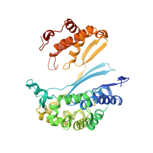
Structure of 6-diazo-5-oxo-norleucine-bound human gamma-glutamyl transpeptidase 1, a novel mechanism of inactivation.

Terzyan, S.S.Cook, P.F.Heroux, A.Hanigan, M.H.

(2017) Protein Sci 26: 1196-1205

  • DOI: https://doi.org/10.1002/pro.3172
  • Primary Citation of Related Structures:  
    5V4Q

  • PubMed Abstract: 

    Intense efforts are underway to identify inhibitors of the enzyme gamma-glutamyl transpeptidase 1 (GGT1) which cleaves extracellular gamma-glutamyl compounds and contributes to the pathology of asthma, reperfusion injury and cancer. The glutamate analog, 6-diazo-5-oxo-norleucine (DON), inhibits GGT1. DON also inhibits many essential glutamine metabolizing enzymes rendering it too toxic for use in the clinic as a GGT1 inhibitor. We investigated the molecular mechanism of human GGT1 (hGGT1) inhibition by DON to determine possible strategies for increasing its specificity for hGGT1. DON is an irreversible inhibitor of hGGT1. The second order rate constant of inactivation was 0.052 mM -1 min -1 and the K i was 2.7 ± 0.7 mM. The crystal structure of DON-inactivated hGGT1 contained a molecule of DON without the diazo-nitrogen atoms in the active site. The overall structure of the hGGT1-DON complex resembled the structure of the apo-enzyme; however, shifts were detected in the loop forming the oxyanion hole and elements of the main chain that form the entrance to the active site. The structure of hGGT1-DON complex revealed two covalent bonds between the enzyme and inhibitor which were part of a six membered ring. The ring included the OG atom of Thr381, the reactive nucleophile of hGGT1 and the α-amine of Thr381. The structure of DON-bound hGGT1 has led to the discovery of a new mechanism of inactivation by DON that differs from its inactivation of other glutamine metabolizing enzymes, and insight into the activation of the catalytic nucleophile that initiates the hGGT1 reaction.


  • Organizational Affiliation

    Laboratory of Biomolecular Structure and Function, University of Oklahoma Health Sciences Center, Oklahoma City, Oklahoma, 73104.


Macromolecules
Find similar proteins by:  (by identity cutoff)  |  3D Structure
Entity ID: 1
MoleculeChains Sequence LengthOrganismDetailsImage
Gamma-glutamyltranspeptidase 1 heavy chain353Homo sapiensMutation(s): 0 
Gene Names: GGT1GGT
EC: 2.3.2.2 (PDB Primary Data), 3.4.19.13 (PDB Primary Data), 3.4.19.14 (PDB Primary Data)
UniProt & NIH Common Fund Data Resources
Find proteins for P19440 (Homo sapiens)
Explore P19440 
Go to UniProtKB:  P19440
PHAROS:  P19440
GTEx:  ENSG00000100031 
Entity Groups  
Sequence Clusters30% Identity50% Identity70% Identity90% Identity95% Identity100% Identity
UniProt GroupP19440
Sequence Annotations
Expand
  • Reference Sequence
Find similar proteins by:  (by identity cutoff)  |  3D Structure
Entity ID: 2
MoleculeChains Sequence LengthOrganismDetailsImage
Gamma-glutamyltranspeptidase 1 light chain189Homo sapiensMutation(s): 0 
Gene Names: GGT1GGT
EC: 2.3.2.2 (PDB Primary Data), 3.4.19.13 (PDB Primary Data), 3.4.19.14 (PDB Primary Data)
UniProt & NIH Common Fund Data Resources
Find proteins for P19440 (Homo sapiens)
Explore P19440 
Go to UniProtKB:  P19440
PHAROS:  P19440
GTEx:  ENSG00000100031 
Entity Groups  
Sequence Clusters30% Identity50% Identity70% Identity90% Identity95% Identity100% Identity
UniProt GroupP19440
Sequence Annotations
Expand
  • Reference Sequence
Small Molecules
Ligands 4 Unique
IDChains Name / Formula / InChI Key2D Diagram3D Interactions
NAG
Query on NAG

Download Ideal Coordinates CCD File 
C [auth A]
D [auth A]
E [auth A]
F [auth A]
G [auth A]
C [auth A],
D [auth A],
E [auth A],
F [auth A],
G [auth A],
J [auth B]
2-acetamido-2-deoxy-beta-D-glucopyranose
C8 H15 N O6
OVRNDRQMDRJTHS-FMDGEEDCSA-N
8WY
Query on 8WY

Download Ideal Coordinates CCD File 
K [auth B]5,5-dihydroxy-L-norleucine
C6 H13 N O4
CRZSOCVDEBRAJD-BYPYZUCNSA-N
CL
Query on CL

Download Ideal Coordinates CCD File 
H [auth A],
L [auth B]
CHLORIDE ION
Cl
VEXZGXHMUGYJMC-UHFFFAOYSA-M
NA
Query on NA

Download Ideal Coordinates CCD File 
I [auth A]SODIUM ION
Na
FKNQFGJONOIPTF-UHFFFAOYSA-N
Experimental Data & Validation

Experimental Data

  • Method: X-RAY DIFFRACTION
  • Resolution: 2.20 Å
  • R-Value Free: 0.219 
  • R-Value Work: 0.163 
  • R-Value Observed: 0.166 
  • Space Group: C 2 2 21
Unit Cell:
Length ( Å )Angle ( ˚ )
a = 105.512α = 90
b = 127.345β = 90
c = 103.069γ = 90
Software Package:
Software NamePurpose
REFMACrefinement
HKL-2000data reduction
HKL-2000data scaling
REFMACphasing

Structure Validation

View Full Validation Report


Ligand Structure Quality Assessment 


Entry History & Funding Information

Deposition Data


Funding OrganizationLocationGrant Number
National Institutes of Health/National Institute of General Medical Sciences (NIH/NIGMS)United StatesP20GM103640
National Science Foundation (NSF, United States)United States092269
National Institutes of Health/National Institute of General Medical Sciences (NIH/NIGMS)United StatesP41RR012408, P41GM103473, P41GM111244
Department of Energy (DOE, United States)United StatesDE-AC02-98CH10886, E-SC0012704

Revision History  (Full details and data files)

  • Version 1.0: 2017-04-19
    Type: Initial release
  • Version 1.1: 2017-06-07
    Changes: Database references
  • Version 1.2: 2017-09-20
    Changes: Author supporting evidence
  • Version 1.3: 2019-11-27
    Changes: Author supporting evidence
  • Version 1.4: 2020-07-29
    Type: Remediation
    Reason: Carbohydrate remediation
    Changes: Data collection, Derived calculations, Refinement description, Structure summary
  • Version 1.5: 2023-10-04
    Changes: Data collection, Database references, Refinement description, Structure summary