4ZRL

Structure of the non canonical Poly(A) polymerase complex GLD-2 - GLD-3


Experimental Data Snapshot

  • Method: X-RAY DIFFRACTION
  • Resolution: 2.28 Å
  • R-Value Free: 0.231 
  • R-Value Work: 0.187 
  • R-Value Observed: 0.189 

wwPDB Validation   3D Report Full Report


This is version 2.0 of the entry. See complete history


Literature

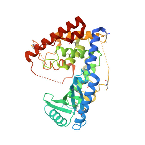
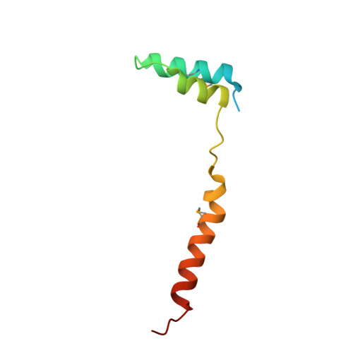
Structural basis for the activation of the C. elegans noncanonical cytoplasmic poly(A)-polymerase GLD-2 by GLD-3.

Nakel, K.Bonneau, F.Eckmann, C.R.Conti, E.

(2015) Proc Natl Acad Sci U S A 112: 8614-8619

  • DOI: https://doi.org/10.1073/pnas.1504648112
  • Primary Citation of Related Structures:  
    4ZRL

  • PubMed Abstract: 

    The Caenorhabditis elegans germ-line development defective (GLD)-2-GLD-3 complex up-regulates the expression of genes required for meiotic progression. GLD-2-GLD-3 acts by extending the short poly(A) tail of germ-line-specific mRNAs, switching them from a dormant state into a translationally active state. GLD-2 is a cytoplasmic noncanonical poly(A) polymerase that lacks the RNA-binding domain typical of the canonical nuclear poly(A)-polymerase Pap1. The activity of C. elegans GLD-2 in vivo and in vitro depends on its association with the multi-K homology (KH) domain-containing protein, GLD-3, a homolog of Bicaudal-C. We have identified a minimal polyadenylation complex that includes the conserved nucleotidyl-transferase core of GLD-2 and the N-terminal domain of GLD-3, and determined its structure at 2.3-Å resolution. The structure shows that the N-terminal domain of GLD-3 does not fold into the predicted KH domain but wraps around the catalytic domain of GLD-2. The picture that emerges from the structural and biochemical data are that GLD-3 activates GLD-2 both indirectly by stabilizing the enzyme and directly by contributing positively charged residues near the RNA-binding cleft. The RNA-binding cleft of GLD-2 has distinct structural features compared with the poly(A)-polymerases Pap1 and Trf4. Consistently, GLD-2 has distinct biochemical properties: It displays unusual specificity in vitro for single-stranded RNAs with at least one adenosine at the 3' end. GLD-2 thus appears to have evolved specialized nucleotidyl-transferase properties that match the 3' end features of dormant cytoplasmic mRNAs.


  • Organizational Affiliation

    Department of Structural Cell Biology, Max Planck Institute of Biochemistry, Am Klopferspitz 18, D-82152 Martinsried, Germany;


Macromolecules
Find similar proteins by:  (by identity cutoff)  |  3D Structure
Entity ID: 1
MoleculeChains Sequence LengthOrganismDetailsImage
Poly(A) RNA polymerase gld-2364Caenorhabditis elegansMutation(s): 0 
Gene Names: gld-2ZC308.1
EC: 2.7.7.19
UniProt
Find proteins for O17087 (Caenorhabditis elegans)
Explore O17087 
Go to UniProtKB:  O17087
Entity Groups  
Sequence Clusters30% Identity50% Identity70% Identity90% Identity95% Identity100% Identity
UniProt GroupO17087
Sequence Annotations
Expand
  • Reference Sequence
Find similar proteins by:  (by identity cutoff)  |  3D Structure
Entity ID: 2
MoleculeChains Sequence LengthOrganismDetailsImage
Defective in germ line development protein 377Caenorhabditis elegansMutation(s): 0 
Gene Names: gld-3T07F8.3
UniProt
Find proteins for Q95ZK7 (Caenorhabditis elegans)
Explore Q95ZK7 
Go to UniProtKB:  Q95ZK7
Entity Groups  
Sequence Clusters30% Identity50% Identity70% Identity90% Identity95% Identity100% Identity
UniProt GroupQ95ZK7
Sequence Annotations
Expand
  • Reference Sequence
Small Molecules
Ligands 1 Unique
IDChains Name / Formula / InChI Key2D Diagram3D Interactions
CL
Query on CL

Download Ideal Coordinates CCD File 
C [auth A]CHLORIDE ION
Cl
VEXZGXHMUGYJMC-UHFFFAOYSA-M
Modified Residues  1 Unique
IDChains TypeFormula2D DiagramParent
MSE
Query on MSE
A
L-PEPTIDE LINKINGC5 H11 N O2 SeMET
Experimental Data & Validation

Experimental Data

  • Method: X-RAY DIFFRACTION
  • Resolution: 2.28 Å
  • R-Value Free: 0.231 
  • R-Value Work: 0.187 
  • R-Value Observed: 0.189 
  • Space Group: P 21 21 21
Unit Cell:
Length ( Å )Angle ( ˚ )
a = 50.655α = 90
b = 89.367β = 90
c = 94.666γ = 90
Software Package:
Software NamePurpose
PHENIXrefinement
XDSdata reduction
SHELXDEphasing
Cootmodel building
XDSdata scaling

Structure Validation

View Full Validation Report


Entry History & Funding Information

Deposition Data


Funding OrganizationLocationGrant Number
German Research FoundationGermanyBU2451/1-2
EC | European Research Council (ERC): Elena Conti ERC Advanced Investigator Grant294371
German Research FoundationSFB646, SFB1035, GRK1721, FOR1680
German Research FoundationFOR855, EC369/3-1

Revision History  (Full details and data files)

  • Version 1.0: 2015-07-08
    Type: Initial release
  • Version 1.1: 2015-07-15
    Changes: Database references
  • Version 1.2: 2015-07-22
    Changes: Database references
  • Version 1.3: 2016-07-13
    Changes: Data collection
  • Version 2.0: 2017-09-06
    Changes: Advisory, Atomic model, Author supporting evidence, Refinement description