3Q6J

Structural basis for carbon dioxide binding by 2-ketopropyl coenzyme M Oxidoreductase/Carboxylase


Experimental Data Snapshot

  • Method: X-RAY DIFFRACTION
  • Resolution: 1.92 Å
  • R-Value Free: 0.221 
  • R-Value Work: 0.164 
  • R-Value Observed: 0.167 

wwPDB Validation   3D Report Full Report


Ligand Structure Quality Assessment 


This is version 1.2 of the entry. See complete history


Literature

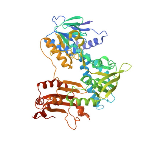
Structural basis for carbon dioxide binding by 2-ketopropyl coenzyme M oxidoreductase/carboxylase.

Pandey, A.S.Mulder, D.W.Ensign, S.A.Peters, J.W.

(2011) FEBS Lett 585: 459-464

  • DOI: https://doi.org/10.1016/j.febslet.2010.12.035
  • Primary Citation of Related Structures:  
    3Q6J

  • PubMed Abstract: 

    The structure of 2-ketopropyl coenzyme M oxidoreductase/carboxylase (2-KPCC) has been determined in a state in which CO(2) is observed providing insights into the mechanism of carboxylation. In the substrate encapsulated state of the enzyme, CO(2) is bound at the base of a narrow hydrophobic substrate access channel. The base of the channel is demarcated by a transition from a hydrophobic to hydrophilic environment where CO(2) is located in position for attack on the carbanion of the ketopropyl group of the substrate to ultimately produce acetoacetate. This binding mode effectively discriminates against H(2)O and prevents protonation of the ketopropyl leaving group.


  • Organizational Affiliation

    Department of Chemistry and Biochemistry, Montana State University, Bozeman, MT 59717, USA.


Macromolecules
Find similar proteins by:  (by identity cutoff)  |  3D Structure
Entity ID: 1
MoleculeChains Sequence LengthOrganismDetailsImage
2-oxopropyl-CoM reductase, carboxylating
A, B
523Xanthobacter autotrophicus Py2Mutation(s): 0 
EC: 1.8.1.5
UniProt
Find proteins for Q56839 (Xanthobacter autotrophicus (strain ATCC BAA-1158 / Py2))
Explore Q56839 
Go to UniProtKB:  Q56839
Entity Groups  
Sequence Clusters30% Identity50% Identity70% Identity90% Identity95% Identity100% Identity
UniProt GroupQ56839
Sequence Annotations
Expand
  • Reference Sequence
Small Molecules
Ligands 8 Unique
IDChains Name / Formula / InChI Key2D Diagram3D Interactions
FAD
Query on FAD

Download Ideal Coordinates CCD File 
D [auth A],
L [auth B]
FLAVIN-ADENINE DINUCLEOTIDE
C27 H33 N9 O15 P2
VWWQXMAJTJZDQX-UYBVJOGSSA-N
NAP
Query on NAP

Download Ideal Coordinates CCD File 
C [auth A],
K [auth B]
NADP NICOTINAMIDE-ADENINE-DINUCLEOTIDE PHOSPHATE
C21 H28 N7 O17 P3
XJLXINKUBYWONI-NNYOXOHSSA-N
KPC
Query on KPC

Download Ideal Coordinates CCD File 
E [auth A](2-[2-KETOPROPYLTHIO]ETHANESULFONATE
C5 H10 O4 S2
CRNXHFXAXBWIRH-UHFFFAOYSA-N
COM
Query on COM

Download Ideal Coordinates CCD File 
M [auth B]1-THIOETHANESULFONIC ACID
C2 H6 O3 S2
ZNEWHQLOPFWXOF-UHFFFAOYSA-N
BCT
Query on BCT

Download Ideal Coordinates CCD File 
G [auth A],
J [auth B]
BICARBONATE ION
C H O3
BVKZGUZCCUSVTD-UHFFFAOYSA-M
ACN
Query on ACN

Download Ideal Coordinates CCD File 
I [auth B]ACETONE
C3 H6 O
CSCPPACGZOOCGX-UHFFFAOYSA-N
CO2
Query on CO2

Download Ideal Coordinates CCD File 
H [auth A]CARBON DIOXIDE
C O2
CURLTUGMZLYLDI-UHFFFAOYSA-N
MG
Query on MG

Download Ideal Coordinates CCD File 
F [auth A],
N [auth B]
MAGNESIUM ION
Mg
JLVVSXFLKOJNIY-UHFFFAOYSA-N
Experimental Data & Validation

Experimental Data

  • Method: X-RAY DIFFRACTION
  • Resolution: 1.92 Å
  • R-Value Free: 0.221 
  • R-Value Work: 0.164 
  • R-Value Observed: 0.167 
  • Space Group: P 1 21 1
Unit Cell:
Length ( Å )Angle ( ˚ )
a = 88.466α = 90
b = 59.803β = 102.28
c = 105.463γ = 90
Software Package:
Software NamePurpose
REFMACrefinement
PDB_EXTRACTdata extraction
MOSFLMdata reduction
SCALAdata scaling
SOLVEphasing

Structure Validation

View Full Validation Report


Ligand Structure Quality Assessment 


Entry History 

Deposition Data

Revision History  (Full details and data files)

  • Version 1.0: 2011-02-16
    Type: Initial release
  • Version 1.1: 2011-07-13
    Changes: Version format compliance
  • Version 1.2: 2023-09-13
    Changes: Data collection, Database references, Derived calculations, Refinement description