3JVF

Crystal structure of an Interleukin-17 receptor complex


Experimental Data Snapshot

  • Method: X-RAY DIFFRACTION
  • Resolution: 3.30 Å
  • R-Value Free: 0.256 
  • R-Value Work: 0.229 
  • R-Value Observed: 0.231 

wwPDB Validation   3D Report Full Report


Ligand Structure Quality Assessment 


This is version 2.0 of the entry. See complete history


Literature

Structural basis of receptor sharing by interleukin 17 cytokines.

Ely, L.K.Fischer, S.Garcia, K.C.

(2009) Nat Immunol 10: 1245-1251

  • DOI: https://doi.org/10.1038/ni.1813
  • Primary Citation of Related Structures:  
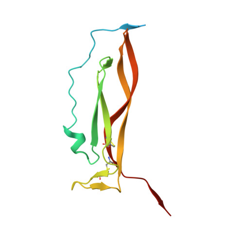
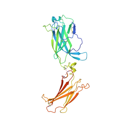
    3JVF

  • PubMed Abstract: 

    Interleukin 17 (IL-17)-producing helper T cells (T(H)-17 cells), together with their effector cytokines, including members of the IL-17 family, are emerging as key mediators of chronic inflammatory and autoimmune disorders. Here we present the crystal structure of a complex of IL-17 receptor A (IL-17RA) bound to IL-17F in a 1:2 stoichiometry. The mechanism of complex formation was unique for cytokines and involved the engagement of IL-17 by two fibronectin-type domains of IL-17RA in a groove between the IL-17 homodimer interface. Binding of the first receptor to the IL-17 cytokines modulated the affinity and specificity of the second receptor-binding event, thereby promoting heterodimeric versus homodimeric complex formation. IL-17RA used a common recognition strategy to bind to several members of the IL-17 family, which allows it to potentially act as a shared receptor in multiple different signaling complexes.


  • Organizational Affiliation

    Howard Hughes Medical Institute, Department of Molecular and Cellular Physiology, Stanford University School of Medicine, Stanford, California, USA.


Macromolecules
Find similar proteins by:  (by identity cutoff)  |  3D Structure
Entity ID: 1
MoleculeChains Sequence LengthOrganismDetailsImage
Interleukin-17F
A, B
133Homo sapiensMutation(s): 0 
Gene Names: IL17FIL24
UniProt & NIH Common Fund Data Resources
Find proteins for Q96PD4 (Homo sapiens)
Explore Q96PD4 
Go to UniProtKB:  Q96PD4
PHAROS:  Q96PD4
GTEx:  ENSG00000112116 
Entity Groups  
Sequence Clusters30% Identity50% Identity70% Identity90% Identity95% Identity100% Identity
UniProt GroupQ96PD4
Sequence Annotations
Expand
  • Reference Sequence
Find similar proteins by:  (by identity cutoff)  |  3D Structure
Entity ID: 2
MoleculeChains Sequence LengthOrganismDetailsImage
Interleukin-17 receptor A286Homo sapiensMutation(s): 0 
Gene Names: IL17RAIL17R
UniProt & NIH Common Fund Data Resources
Find proteins for Q96F46 (Homo sapiens)
Explore Q96F46 
Go to UniProtKB:  Q96F46
PHAROS:  Q96F46
GTEx:  ENSG00000177663 
Entity Groups  
Sequence Clusters30% Identity50% Identity70% Identity90% Identity95% Identity100% Identity
UniProt GroupQ96F46
Sequence Annotations
Expand
  • Reference Sequence
Oligosaccharides

Help

Entity ID: 3
MoleculeChains Length2D Diagram Glycosylation3D Interactions
2-acetamido-2-deoxy-beta-D-glucopyranose-(1-4)-2-acetamido-2-deoxy-beta-D-glucopyranose
D
2N-Glycosylation
Glycosylation Resources
GlyTouCan:  G42666HT
GlyCosmos:  G42666HT
GlyGen:  G42666HT
Experimental Data & Validation

Experimental Data

  • Method: X-RAY DIFFRACTION
  • Resolution: 3.30 Å
  • R-Value Free: 0.256 
  • R-Value Work: 0.229 
  • R-Value Observed: 0.231 
  • Space Group: P 41 21 2
Unit Cell:
Length ( Å )Angle ( ˚ )
a = 170.73α = 90
b = 170.73β = 90
c = 81.9γ = 90
Software Package:
Software NamePurpose
Blu-Icedata collection
SHARPphasing
PHENIXrefinement
MOSFLMdata reduction
SCALAdata scaling

Structure Validation

View Full Validation Report


Ligand Structure Quality Assessment 


Entry History 

Deposition Data

Revision History  (Full details and data files)

  • Version 1.0: 2009-10-20
    Type: Initial release
  • Version 1.1: 2011-07-13
    Changes: Non-polymer description, Version format compliance
  • Version 2.0: 2020-07-29
    Type: Remediation
    Reason: Carbohydrate remediation
    Changes: Advisory, Atomic model, Data collection, Derived calculations, Structure summary