3GV8

Human DNA polymerase iota in complex with T template DNA and incoming dGTP


Experimental Data Snapshot

  • Method: X-RAY DIFFRACTION
  • Resolution: 2.00 Å
  • R-Value Free: 0.248 
  • R-Value Work: 0.218 
  • R-Value Observed: 0.219 

wwPDB Validation   3D Report Full Report


Ligand Structure Quality Assessment 


This is version 1.3 of the entry. See complete history


Literature

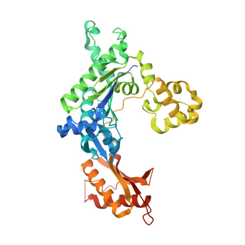
Structural basis of error-prone replication and stalling at a thymine base by human DNA polymerase iota

Kirouac, K.N.Ling, H.

(2009) EMBO J 28: 1644-1654

  • DOI: https://doi.org/10.1038/emboj.2009.122
  • Primary Citation of Related Structures:  
    3GV5, 3GV7, 3GV8

  • PubMed Abstract: 

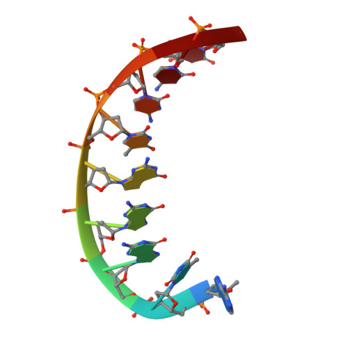
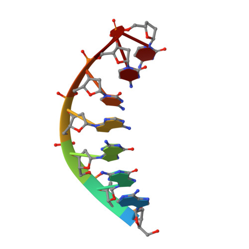
    Human DNA polymerase iota (pol iota) is a unique member of Y-family polymerases, which preferentially misincorporates nucleotides opposite thymines (T) and halts replication at T bases. The structural basis of the high error rates remains elusive. We present three crystal structures of pol complexed with DNA containing a thymine base, paired with correct or incorrect incoming nucleotides. A narrowed active site supports a pyrimidine to pyrimidine mismatch and excludes Watson-Crick base pairing by pol. The template thymine remains in an anti conformation irrespective of incoming nucleotides. Incoming ddATP adopts a syn conformation with reduced base stacking, whereas incorrect dGTP and dTTP maintain anti conformations with normal base stacking. Further stabilization of dGTP by H-bonding with Gln59 of the finger domain explains the preferential T to G mismatch. A template 'U-turn' is stabilized by pol and the methyl group of the thymine template, revealing the structural basis of T stalling. Our structural and domain-swapping experiments indicate that the finger domain is responsible for pol's high error rates on pyrimidines and determines the incorporation specificity.


  • Organizational Affiliation

    Department of Biochemistry, University of Western Ontario, London, Ontario, Canada.


Macromolecules

Find similar proteins by:  (by identity cutoff)  |  3D Structure
Entity ID: 1
MoleculeChains Sequence LengthOrganismDetailsImage
DNA polymerase iotaA [auth B]420Homo sapiensMutation(s): 0 
Gene Names: POLIRAD30B
EC: 2.7.7.7
UniProt & NIH Common Fund Data Resources
Find proteins for Q9UNA4 (Homo sapiens)
Explore Q9UNA4 
Go to UniProtKB:  Q9UNA4
PHAROS:  Q9UNA4
GTEx:  ENSG00000101751 
Entity Groups  
Sequence Clusters30% Identity50% Identity70% Identity90% Identity95% Identity100% Identity
UniProt GroupQ9UNA4
Sequence Annotations
Expand
  • Reference Sequence

Find similar nucleic acids by:  Sequence   |   3D Structure  

Entity ID: 2
MoleculeChains LengthOrganismImage
5'-D(*AP*TP*GP*GP*GP*TP*CP*CP*T)-3'B [auth T]9N/A
Sequence Annotations
Expand
  • Reference Sequence

Find similar nucleic acids by:  Sequence   |   3D Structure  

Entity ID: 3
MoleculeChains LengthOrganismImage
5'-D(*AP*GP*GP*AP*CP*CP*C)-3'C [auth P]7N/A
Sequence Annotations
Expand
  • Reference Sequence
Experimental Data & Validation

Experimental Data

  • Method: X-RAY DIFFRACTION
  • Resolution: 2.00 Å
  • R-Value Free: 0.248 
  • R-Value Work: 0.218 
  • R-Value Observed: 0.219 
  • Space Group: P 65 2 2
Unit Cell:
Length ( Å )Angle ( ˚ )
a = 98.07α = 90
b = 98.07β = 90
c = 203.38γ = 120
Software Package:
Software NamePurpose
DENZOdata reduction
SCALEPACKdata scaling
REFMACrefinement
PDB_EXTRACTdata extraction

Structure Validation

View Full Validation Report


Ligand Structure Quality Assessment 


Entry History 

Deposition Data

Revision History  (Full details and data files)

  • Version 1.0: 2009-06-02
    Type: Initial release
  • Version 1.1: 2011-07-13
    Changes: Advisory, Version format compliance
  • Version 1.2: 2017-11-01
    Changes: Refinement description
  • Version 1.3: 2024-02-21
    Changes: Data collection, Database references, Derived calculations