3B9X

Crystal structure of the E. coli pyrimidine nucleoside hydrolase YeiK in complex with inosine


Experimental Data Snapshot

  • Method: X-RAY DIFFRACTION
  • Resolution: 2.30 Å
  • R-Value Free: 0.255 
  • R-Value Work: 0.201 
  • R-Value Observed: 0.203 

wwPDB Validation   3D Report Full Report


Ligand Structure Quality Assessment 


This is version 1.2 of the entry. See complete history


Literature

Structural basis for substrate specificity in group I nucleoside hydrolases

Iovane, E.Giabbai, B.Muzzolini, L.Matafora, V.Fornili, A.Minici, C.Giannese, F.Degano, M.

(2008) Biochemistry 47: 4418-4426

  • DOI: https://doi.org/10.1021/bi702448s
  • Primary Citation of Related Structures:  
    3B9X

  • PubMed Abstract: 

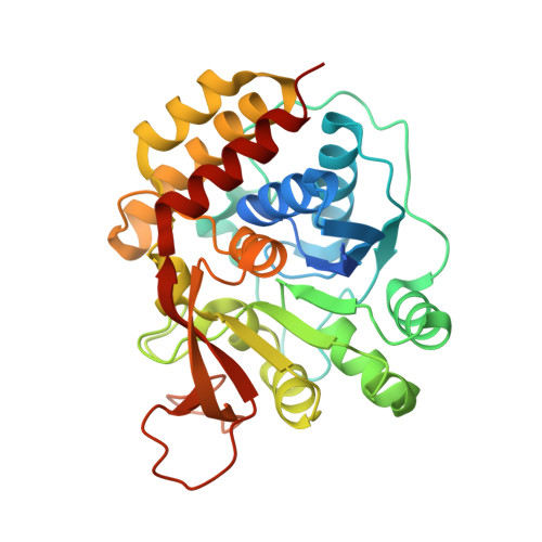
    Enzymes with nucleoside hydrolase activity (NHs) belonging to homology group I either are markedly specific for pyrimidine nucleoside substrates or hydrolyze with comparable efficiencies the N-glycosidic bond in all common nucleosides. The biochemical and structural basis for these differences in substrate specificity is still unknown. Here we characterize the binding interactions between the slowly hydrolyzed substrate inosine and the Escherichia coli pyrimidine-specific NH YeiK using cryotrapping and X-ray crystallography. Guided by the structural features of the Michaelis complex, we show the synergic effect of two specific point mutations in YeiK that increase the catalytic efficiency toward purine nucleosides to values comparable to those of natural nonspecific NHs. We demonstrate that the integrity of an active-site catalytic triad comprised of two hydroxylated amino acids and one histidine residue is a requirement for the highly efficient hydrolysis of inosine by group I NHs. Instead, cleavage of the YeiK-preferred substrate uridine is not affected by mutations at the same locations, suggesting a different fine chemical mechanism for the hydrolysis of the two nucleoside substrates. Our study provides for the first time direct evidence that distinct subsets of amino acid residues are involved in the hydrolysis of purine or pyrimidine nucleosides in group I NHs.


  • Organizational Affiliation

    Biocrystallography Unit, DIBIT San Raffaele Scientific Institute, via Olgettina 58, 20132 Milan, Italy.


Macromolecules
Find similar proteins by:  (by identity cutoff)  |  3D Structure
Entity ID: 1
MoleculeChains Sequence LengthOrganismDetailsImage
Pyrimidine-specific ribonucleoside hydrolase rihB
A, B, C, D
333Escherichia coli K-12Mutation(s): 0 
Gene Names: rihByeiK
EC: 3.2.2.8
UniProt
Find proteins for P33022 (Escherichia coli (strain K12))
Explore P33022 
Go to UniProtKB:  P33022
Entity Groups  
Sequence Clusters30% Identity50% Identity70% Identity90% Identity95% Identity100% Identity
UniProt GroupP33022
Sequence Annotations
Expand
  • Reference Sequence
Small Molecules
Ligands 3 Unique
IDChains Name / Formula / InChI Key2D Diagram3D Interactions
NOS
Query on NOS

Download Ideal Coordinates CCD File 
F [auth A],
H [auth B],
J [auth C],
L [auth D]
INOSINE
C10 H12 N4 O5
UGQMRVRMYYASKQ-KQYNXXCUSA-N
TAM
Query on TAM

Download Ideal Coordinates CCD File 
M [auth D]TRIS(HYDROXYETHYL)AMINOMETHANE
C7 H17 N O3
GKODZWOPPOTFGA-UHFFFAOYSA-N
CA
Query on CA

Download Ideal Coordinates CCD File 
E [auth A],
G [auth B],
I [auth C],
K [auth D]
CALCIUM ION
Ca
BHPQYMZQTOCNFJ-UHFFFAOYSA-N
Experimental Data & Validation

Experimental Data

  • Method: X-RAY DIFFRACTION
  • Resolution: 2.30 Å
  • R-Value Free: 0.255 
  • R-Value Work: 0.201 
  • R-Value Observed: 0.203 
  • Space Group: P 1
Unit Cell:
Length ( Å )Angle ( ˚ )
a = 45.112α = 67.9
b = 82.406β = 79.59
c = 90.163γ = 89.65
Software Package:
Software NamePurpose
REFMACrefinement
CrystalCleardata collection
XDSdata reduction
XSCALEdata scaling
MOLREPphasing

Structure Validation

View Full Validation Report


Ligand Structure Quality Assessment 


Entry History 

Deposition Data

Revision History  (Full details and data files)

  • Version 1.0: 2008-04-01
    Type: Initial release
  • Version 1.1: 2011-07-13
    Changes: Source and taxonomy, Version format compliance
  • Version 1.2: 2023-11-01
    Changes: Data collection, Database references, Derived calculations, Refinement description