2OJQ

Crystal structure of Alix V domain


Experimental Data Snapshot

  • Method: X-RAY DIFFRACTION
  • Resolution: 2.87 Å
  • R-Value Free: 0.318 
  • R-Value Work: 0.250 
  • R-Value Observed: 0.257 

wwPDB Validation   3D Report Full Report


This is version 1.4 of the entry. See complete history


Literature

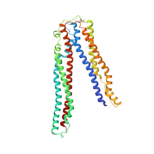
Structural basis for viral late-domain binding to Alix

Lee, S.Joshi, A.Nagashima, K.Freed, E.O.Hurley, J.H.

(2007) Nat Struct Mol Biol 14: 194-199

  • DOI: https://doi.org/10.1038/nsmb1203
  • Primary Citation of Related Structures:  
    2OJQ

  • PubMed Abstract: 

    The modular protein Alix is a central node in endosomal-lysosomal trafficking and the budding of human immunodeficiency virus (HIV)-1. The Gag p6 protein of HIV-1 contains a LYPx(n)LxxL motif that is required for Alix-mediated budding and binds a region of Alix spanning residues 360-702. The structure of this fragment of Alix has the shape of the letter 'V' and is termed the V domain. The V domain has a topologically complex arrangement of 11 alpha-helices, with connecting loops that cross three times between the two arms of the V. The conserved residue Phe676 is at the center of a large hydrophobic pocket and is crucial for binding to a peptide model of HIV-1 p6. Overexpression of the V domain inhibits HIV-1 release from cells. This inhibition of release is reversed by mutations that block binding of the Alix V domain to p6.


  • Organizational Affiliation

    Laboratory of Molecular Biology, National Institute of Diabetes and Digestive and Kidney Diseases, National Institutes of Health (NIH), US Department of Health and Human Services, Bethesda, Maryland 20892, USA.


Macromolecules
Find similar proteins by:  (by identity cutoff)  |  3D Structure
Entity ID: 1
MoleculeChains Sequence LengthOrganismDetailsImage
Programmed cell death 6-interacting protein348Homo sapiensMutation(s): 4 
Gene Names: PDCD6IPAIP1ALIXKIAA1375
UniProt & NIH Common Fund Data Resources
Find proteins for Q8WUM4 (Homo sapiens)
Explore Q8WUM4 
Go to UniProtKB:  Q8WUM4
PHAROS:  Q8WUM4
GTEx:  ENSG00000170248 
Entity Groups  
Sequence Clusters30% Identity50% Identity70% Identity90% Identity95% Identity100% Identity
UniProt GroupQ8WUM4
Sequence Annotations
Expand
  • Reference Sequence
Small Molecules
Modified Residues  1 Unique
IDChains TypeFormula2D DiagramParent
MSE
Query on MSE
A
L-PEPTIDE LINKINGC5 H11 N O2 SeMET
Experimental Data & Validation

Experimental Data

  • Method: X-RAY DIFFRACTION
  • Resolution: 2.87 Å
  • R-Value Free: 0.318 
  • R-Value Work: 0.250 
  • R-Value Observed: 0.257 
  • Space Group: P 1 21 1
Unit Cell:
Length ( Å )Angle ( ˚ )
a = 37.65α = 90
b = 55.38β = 92.37
c = 113.166γ = 90
Software Package:
Software NamePurpose
REFMACrefinement
HKL-2000data collection
HKL-2000data reduction
HKL-2000data scaling
SOLVEphasing

Structure Validation

View Full Validation Report


Entry History 

Deposition Data

Revision History  (Full details and data files)

  • Version 1.0: 2007-02-20
    Type: Initial release
  • Version 1.1: 2008-05-01
    Changes: Version format compliance
  • Version 1.2: 2011-07-13
    Changes: Advisory, Version format compliance
  • Version 1.3: 2018-01-31
    Changes: Experimental preparation
  • Version 1.4: 2023-12-27
    Changes: Data collection, Database references, Derived calculations