1LDP

CRYSTAL STRUCTURE OF MURINE MHC CLASS I H-2LD WITH A MIXTURE OF BOUND PEPTIDES


Experimental Data Snapshot

  • Method: X-RAY DIFFRACTION
  • Resolution: 3.10 Å
  • R-Value Free: 0.371 
  • R-Value Work: 0.221 
  • R-Value Observed: 0.221 

wwPDB Validation   3D Report Full Report


This is version 2.1 of the entry. See complete history


Literature

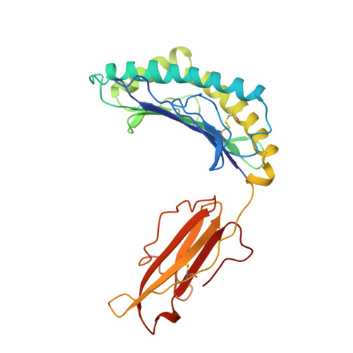
Structural basis of 2C TCR allorecognition of H-2Ld peptide complexes.

Speir, J.A.Garcia, K.C.Brunmark, A.Degano, M.Peterson, P.A.Teyton, L.Wilson, I.A.

(1998) Immunity 8: 553-562

  • DOI: https://doi.org/10.1016/s1074-7613(00)80560-9
  • Primary Citation of Related Structures:  
    1LDP

  • PubMed Abstract: 

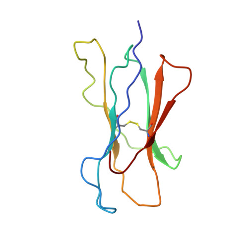
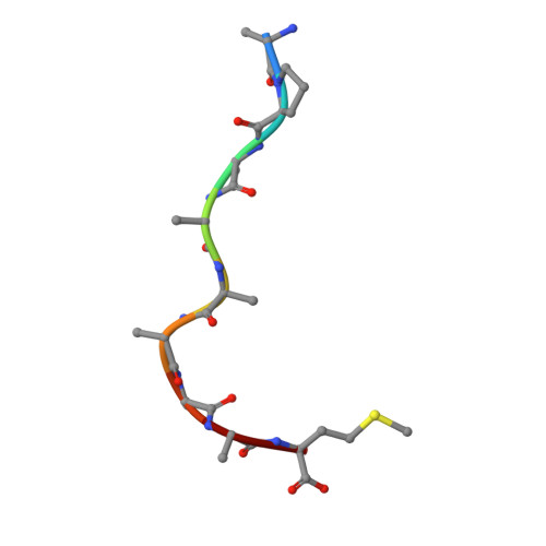
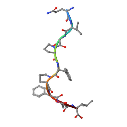
    MHC class I H-2Ld complexed with peptide QL9 (or p2Ca) is a high-affinity alloantigen for the 2C TCR. We used the crystal structure of H-2Ld with a mixture of bound peptides at 3.1 A to construct a model of the allogeneic 2C-Ld/QL9 complex for comparison with the syngeneic 2C-Kb/dEV8 structure. A prominent ridge on the floor of the Ld peptide-binding groove, not present in Kb, creates a C-terminal bulge in Ld peptides that greatly increases interactions with the 2C beta-chain. Furthermore, weak electrostatic complementarity between Asp77 on the alpha1 helix of Kb and 2C is enhanced in the allogeneic complex by closer proximity of QL9 peptide residue AspP8 to the 2C HV4 loop.


  • Organizational Affiliation

    Department of Molecular Biology, The Scripps Research Institute, La Jolla, California 92037, USA.


Macromolecules
Find similar proteins by:  (by identity cutoff)  |  3D Structure
Entity ID: 1
MoleculeChains Sequence LengthOrganismDetailsImage
MHC CLASS I H-2LDA [auth H]272Mus musculusMutation(s): 0 
Gene Names: H-2LD
UniProt
Find proteins for P01897 (Mus musculus)
Explore P01897 
Go to UniProtKB:  P01897
Entity Groups  
Sequence Clusters30% Identity50% Identity70% Identity90% Identity95% Identity100% Identity
UniProt GroupP01897
Sequence Annotations
Expand
  • Reference Sequence
Find similar proteins by:  (by identity cutoff)  |  3D Structure
Entity ID: 2
MoleculeChains Sequence LengthOrganismDetailsImage
MHC CLASS I H-2LDB [auth L]99Mus musculusMutation(s): 0 
Gene Names: H-2LD
UniProt & NIH Common Fund Data Resources
Find proteins for P01887 (Mus musculus)
Explore P01887 
Go to UniProtKB:  P01887
IMPC:  MGI:88127
Entity Groups  
Sequence Clusters30% Identity50% Identity70% Identity90% Identity95% Identity100% Identity
UniProt GroupP01887
Sequence Annotations
Expand
  • Reference Sequence

Find similar proteins by:  Sequence   |   3D Structure  

Entity ID: 3
MoleculeChains Sequence LengthOrganismDetailsImage
PEPTIDEC [auth P]9Drosophila melanogasterMutation(s): 0 
Sequence Annotations
Expand
  • Reference Sequence

Find similar proteins by:  Sequence   |   3D Structure  

Entity ID: 4
MoleculeChains Sequence LengthOrganismDetailsImage
PEPTIDED [auth Q]9Drosophila melanogasterMutation(s): 0 
Sequence Annotations
Expand
  • Reference Sequence
Oligosaccharides

Help

Entity ID: 5
MoleculeChains Length2D Diagram Glycosylation3D Interactions
2-acetamido-2-deoxy-alpha-D-glucopyranose-(1-4)-2-acetamido-2-deoxy-beta-D-glucopyranoseE [auth A]2N-Glycosylation
Glycosylation Resources
GlyTouCan:  G07375KG
GlyCosmos:  G07375KG
GlyGen:  G07375KG
Small Molecules
Ligands 1 Unique
IDChains Name / Formula / InChI Key2D Diagram3D Interactions
NAG
Query on NAG

Download Ideal Coordinates CCD File 
F [auth H]2-acetamido-2-deoxy-beta-D-glucopyranose
C8 H15 N O6
OVRNDRQMDRJTHS-FMDGEEDCSA-N
Experimental Data & Validation

Experimental Data

  • Method: X-RAY DIFFRACTION
  • Resolution: 3.10 Å
  • R-Value Free: 0.371 
  • R-Value Work: 0.221 
  • R-Value Observed: 0.221 
  • Space Group: P 1 2 1
Unit Cell:
Length ( Å )Angle ( ˚ )
a = 72.4α = 90
b = 48.98β = 97.95
c = 84.06γ = 90
Software Package:
Software NamePurpose
XDSdata scaling
SCALEPACKdata scaling
X-PLORmodel building
X-PLORrefinement
XDSdata reduction
X-PLORphasing

Structure Validation

View Full Validation Report


Entry History 

Deposition Data

Revision History  (Full details and data files)

  • Version 1.0: 1998-06-17
    Type: Initial release
  • Version 1.1: 2008-03-03
    Changes: Version format compliance
  • Version 1.2: 2011-07-13
    Changes: Version format compliance
  • Version 2.0: 2020-07-29
    Type: Remediation
    Reason: Carbohydrate remediation
    Changes: Advisory, Atomic model, Data collection, Derived calculations, Non-polymer description, Structure summary
  • Version 2.1: 2023-08-09
    Changes: Database references, Refinement description, Structure summary