6L8Q

Complex structure of bat CD26 and MERS-RBD


Experimental Data Snapshot

  • Method: X-RAY DIFFRACTION
  • Resolution: 3.10 Å
  • R-Value Free: 0.241 
  • R-Value Work: 0.200 
  • R-Value Observed: 0.202 

wwPDB Validation   3D Report Full Report


Ligand Structure Quality Assessment 


This is version 2.1 of the entry. See complete history


Literature

Molecular Basis of Binding between Middle East Respiratory Syndrome Coronavirus and CD26 from Seven Bat Species.

Yuan, Y.Qi, J.Peng, R.Li, C.Lu, G.Yan, J.Wang, Q.Gao, G.F.

(2020) J Virol 94

  • DOI: https://doi.org/10.1128/JVI.01387-19
  • Primary Citation of Related Structures:  
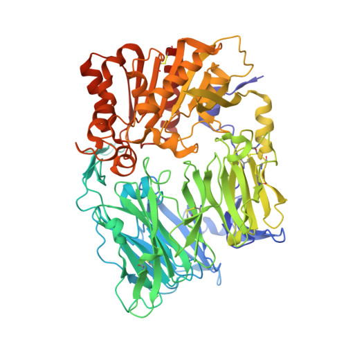
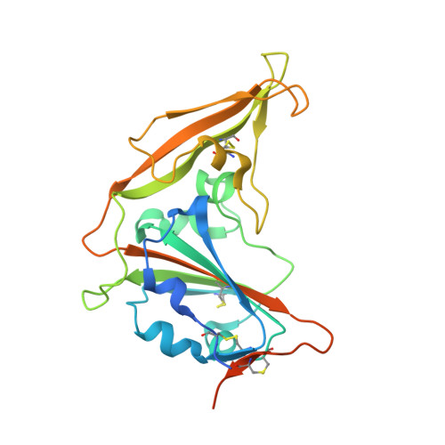
    6L8Q

  • PubMed Abstract: 

    Continued reports of Middle East respiratory syndrome coronavirus (MERS-CoV) infecting humans have occurred since the identification of this virus in 2012. MERS-CoV is prone to cause endemic disease in the Middle East, with several dozen spillover infections to other continents. It is hypothesized that MERS-CoV originated from bat coronaviruses and that dromedary camels are its natural reservoir. Although gene segments identical to MERS-CoV were sequenced from certain species of bats and one species experimentally shed the virus, it is still unknown whether other bats can transmit the virus. Here, at the molecular level, we found that all purified bat CD26s (bCD26s) from a diverse range of species interact with the receptor binding domain (RBD) of MERS-CoV, with equilibrium dissociation constant values ranging from several to hundreds at the micromolar level. Moreover, all bCD26s expressed in this study mediated the entry of pseudotyped MERS-CoV to receptor-expressing cells, indicating the broad potential engagement of bCD26s as MERS-CoV receptors. Further structural analysis indicated that in the bat receptor, compared to the human receptor, substitutions of key residues and their adjacent amino acids leads to decreased binding affinity to the MERS-RBD. These results add more evidence to the existing belief that bats are the original source of MERS-CoV and suggest that bCD26s in many species can mediate the entry of the virus, which has significant implications for the surveillance and control of MERS-CoV infection. IMPORTANCE In this study, we found that bat CD26s (bCD26s) from different species exhibit large diversities, especially in the region responsible for binding to the receptor binding domain (RBD) of Middle East respiratory syndrome coronavirus (MERS-CoV). However, they maintain the interaction with MERS-RBD at varied affinities and support the entry of pseudotyped MERS-CoV. These bat receptors polymorphisms seem to confer evolutionary pressure for the adaptation of CD26-binding virus, such as the ancestor of MERS-CoV, and led to the generation of diversified CD26-engaging CoV strains. Thus, our data add more evidence to support that bats are the reservoir of MERS-CoV and similar viruses, as well as further emphasize the necessity to survey MERS-CoV and other CoVs among bats.


  • Organizational Affiliation

    CAS Key Laboratory of Pathogenic Microbiology and Immunology, Institute of Microbiology, Chinese Academy of Sciences, Beijing, China.


Macromolecules
Find similar proteins by:  (by identity cutoff)  |  3D Structure
Entity ID: 1
MoleculeChains Sequence LengthOrganismDetailsImage
Dipeptidyl peptidase 4
A, C, E, G
731Myotis davidiiMutation(s): 0 
Gene Names: MDA_GLEAN10024208
UniProt
Find proteins for L5LQ33 (Myotis davidii)
Explore L5LQ33 
Go to UniProtKB:  L5LQ33
Entity Groups  
Sequence Clusters30% Identity50% Identity70% Identity90% Identity95% Identity100% Identity
UniProt GroupL5LQ33
Sequence Annotations
Expand
  • Reference Sequence
Find similar proteins by:  (by identity cutoff)  |  3D Structure
Entity ID: 2
MoleculeChains Sequence LengthOrganismDetailsImage
Spike glycoprotein
B, D, F, H
246Middle East respiratory syndrome-related coronavirusMutation(s): 0 
UniProt
Find proteins for K9N5Q8 (Middle East respiratory syndrome-related coronavirus (isolate United Kingdom/H123990006/2012))
Explore K9N5Q8 
Go to UniProtKB:  K9N5Q8
Entity Groups  
Sequence Clusters30% Identity50% Identity70% Identity90% Identity95% Identity100% Identity
UniProt GroupK9N5Q8
Sequence Annotations
Expand
  • Reference Sequence
Oligosaccharides

Help

Entity ID: 3
MoleculeChains Length2D Diagram Glycosylation3D Interactions
2-acetamido-2-deoxy-beta-D-glucopyranose-(1-4)-2-acetamido-2-deoxy-beta-D-glucopyranose
I, K, L, M, O
I, K, L, M, O, P, Q, R, T, U, V, X, Y
2N-Glycosylation
Glycosylation Resources
GlyTouCan:  G42666HT
GlyCosmos:  G42666HT
GlyGen:  G42666HT
Entity ID: 4
MoleculeChains Length2D Diagram Glycosylation3D Interactions
beta-D-mannopyranose-(1-4)-2-acetamido-2-deoxy-beta-D-glucopyranose-(1-4)-2-acetamido-2-deoxy-beta-D-glucopyranose
J, N, S, W
3N-Glycosylation
Glycosylation Resources
GlyTouCan:  G15407YE
GlyCosmos:  G15407YE
GlyGen:  G15407YE
Small Molecules
Ligands 1 Unique
IDChains Name / Formula / InChI Key2D Diagram3D Interactions
NAG
Query on NAG

Download Ideal Coordinates CCD File 
AA [auth A]
BA [auth B]
CA [auth C]
DA [auth C]
EA [auth D]
AA [auth A],
BA [auth B],
CA [auth C],
DA [auth C],
EA [auth D],
FA [auth E],
GA [auth F],
HA [auth G],
IA [auth G],
JA [auth H],
Z [auth A]
2-acetamido-2-deoxy-beta-D-glucopyranose
C8 H15 N O6
OVRNDRQMDRJTHS-FMDGEEDCSA-N
Experimental Data & Validation

Experimental Data

  • Method: X-RAY DIFFRACTION
  • Resolution: 3.10 Å
  • R-Value Free: 0.241 
  • R-Value Work: 0.200 
  • R-Value Observed: 0.202 
  • Space Group: P 1 21 1
Unit Cell:
Length ( Å )Angle ( ˚ )
a = 114.657α = 90
b = 273.674β = 119.682
c = 115.234γ = 90
Software Package:
Software NamePurpose
PHENIXrefinement
HKL-2000data reduction
SCALEPACKdata scaling
PHASERphasing

Structure Validation

View Full Validation Report


Ligand Structure Quality Assessment 


Entry History & Funding Information

Deposition Data

  • Released Date: 2019-12-04 
  • Deposition Author(s): Yuan, Y.

Funding OrganizationLocationGrant Number
National Science Foundation (China)China--

Revision History  (Full details and data files)

  • Version 1.0: 2019-12-04
    Type: Initial release
  • Version 1.1: 2020-06-17
    Changes: Data collection, Database references
  • Version 2.0: 2020-07-29
    Type: Remediation
    Reason: Carbohydrate remediation
    Changes: Atomic model, Data collection, Derived calculations, Structure summary
  • Version 2.1: 2023-11-22
    Changes: Data collection, Database references, Refinement description, Structure summary