6AHY

Wnt signaling complex


Experimental Data Snapshot

  • Method: X-RAY DIFFRACTION
  • Resolution: 2.80 Å
  • R-Value Free: 0.244 
  • R-Value Work: 0.205 
  • R-Value Observed: 0.207 

wwPDB Validation   3D Report Full Report


Ligand Structure Quality Assessment 


This is version 2.1 of the entry. See complete history


Literature

Crystal structure of a mammalian Wnt-frizzled complex.

Hirai, H.Matoba, K.Mihara, E.Arimori, T.Takagi, J.

(2019) Nat Struct Mol Biol 26: 372-379

  • DOI: https://doi.org/10.1038/s41594-019-0216-z
  • Primary Citation of Related Structures:  
    6AHY

  • PubMed Abstract: 

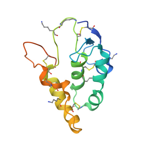
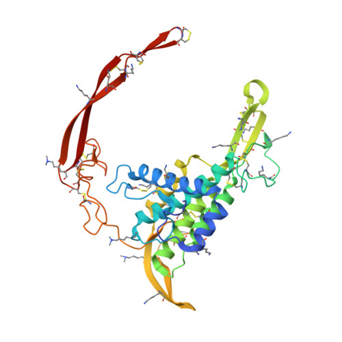
    Wnt signaling plays fundamental roles in organogenesis, tissue regeneration and cancer, but high-resolution structural information of mammalian Wnt proteins is lacking. We solved a 2.8-Å resolution crystal structure of human Wnt3 in complex with mouse Frizzled 8 Cys-rich domain (CRD). Wnt3 grabs the receptor in a manner very similar to that found in Xenopus Wnt8 complexed with the same receptor. Unlike Xenopus Wnt8-bound CRD, however, Wnt3-bound CRD formed a symmetrical dimer in the crystal by exchanging the tip of the unsaturated acyl chain attached to each Wnt3, confirming the ability of Wnt and Frizzled CRD to form a 2:2 complex. The hypervariable 'linker' region of Wnt3 formed a β-hairpin protrusion opposite from the Frizzled binding interface, consistent with its proposed role in the coreceptor recognition. Direct binding between this segment and the Wnt coreceptor LRP6 was confirmed, enabling us to build a structural model of the Wnt-Frizzled-LRP6 ternary complex.


  • Organizational Affiliation

    Laboratory of Protein Synthesis and Expression, Institute for Protein Research, Osaka University, Osaka, Japan.


Macromolecules
Find similar proteins by:  (by identity cutoff)  |  3D Structure
Entity ID: 1
MoleculeChains Sequence LengthOrganismDetailsImage
Frizzled-8
A, C, E
141Mus musculusMutation(s): 0 
Gene Names: Fzd8
UniProt & NIH Common Fund Data Resources
Find proteins for Q61091 (Mus musculus)
Explore Q61091 
Go to UniProtKB:  Q61091
IMPC:  MGI:108460
Entity Groups  
Sequence Clusters30% Identity50% Identity70% Identity90% Identity95% Identity100% Identity
UniProt GroupQ61091
Sequence Annotations
Expand
  • Reference Sequence
Find similar proteins by:  (by identity cutoff)  |  3D Structure
Entity ID: 2
MoleculeChains Sequence LengthOrganismDetailsImage
Proto-oncogene Wnt-3
B, D, F
319Homo sapiensMutation(s): 0 
Gene Names: WNT3INT4
UniProt & NIH Common Fund Data Resources
Find proteins for P56703 (Homo sapiens)
Explore P56703 
Go to UniProtKB:  P56703
PHAROS:  P56703
GTEx:  ENSG00000108379 
Entity Groups  
Sequence Clusters30% Identity50% Identity70% Identity90% Identity95% Identity100% Identity
UniProt GroupP56703
Sequence Annotations
Expand
  • Reference Sequence
Oligosaccharides

Help

Entity ID: 3
MoleculeChains Length2D Diagram Glycosylation3D Interactions
2-acetamido-2-deoxy-beta-D-glucopyranose-(1-4)-2-acetamido-2-deoxy-beta-D-glucopyranose
G, H
2N-Glycosylation
Glycosylation Resources
GlyTouCan:  G42666HT
GlyCosmos:  G42666HT
GlyGen:  G42666HT
Small Molecules
Ligands 3 Unique
IDChains Name / Formula / InChI Key2D Diagram3D Interactions
PAM
Query on PAM

Download Ideal Coordinates CCD File 
L [auth B],
O [auth D]
PALMITOLEIC ACID
C16 H30 O2
SECPZKHBENQXJG-FPLPWBNLSA-N
NAG
Query on NAG

Download Ideal Coordinates CCD File 
I [auth A]
K [auth B]
M [auth C]
N [auth D]
P [auth E]
I [auth A],
K [auth B],
M [auth C],
N [auth D],
P [auth E],
Q [auth F]
2-acetamido-2-deoxy-beta-D-glucopyranose
C8 H15 N O6
OVRNDRQMDRJTHS-FMDGEEDCSA-N
CL
Query on CL

Download Ideal Coordinates CCD File 
J [auth B]CHLORIDE ION
Cl
VEXZGXHMUGYJMC-UHFFFAOYSA-M
Modified Residues  1 Unique
IDChains TypeFormula2D DiagramParent
MLY
Query on MLY
A, C, E
L-PEPTIDE LINKINGC8 H18 N2 O2LYS
Experimental Data & Validation

Experimental Data

  • Method: X-RAY DIFFRACTION
  • Resolution: 2.80 Å
  • R-Value Free: 0.244 
  • R-Value Work: 0.205 
  • R-Value Observed: 0.207 
  • Space Group: I 21 21 21
Unit Cell:
Length ( Å )Angle ( ˚ )
a = 120.073α = 90
b = 141.611β = 90
c = 260.692γ = 90
Software Package:
Software NamePurpose
PHENIXrefinement
HKL-2000data reduction
HKL-2000data scaling
PHASERphasing

Structure Validation

View Full Validation Report


Ligand Structure Quality Assessment 


Entry History 

Deposition Data

Revision History  (Full details and data files)

  • Version 1.0: 2019-05-01
    Type: Initial release
  • Version 1.1: 2019-05-15
    Changes: Data collection, Database references
  • Version 1.2: 2019-05-22
    Changes: Data collection, Database references
  • Version 2.0: 2020-07-29
    Type: Remediation
    Reason: Carbohydrate remediation
    Changes: Advisory, Atomic model, Data collection, Derived calculations, Structure summary
  • Version 2.1: 2023-11-22
    Changes: Data collection, Database references, Refinement description, Structure summary