6NC3

AMC011 v4.2 SOSIP Env trimer in complex with fusion peptide targeting antibody VRC34 fragment antigen binding


Experimental Data Snapshot

  • Method: ELECTRON MICROSCOPY
  • Resolution: 4.50 Å
  • Aggregation State: PARTICLE 
  • Reconstruction Method: SINGLE PARTICLE 

wwPDB Validation   3D Report Full Report


This is version 2.0 of the entry. See complete history


Literature

Conformational Plasticity in the HIV-1 Fusion Peptide Facilitates Recognition by Broadly Neutralizing Antibodies.

Yuan, M.Cottrell, C.A.Ozorowski, G.van Gils, M.J.Kumar, S.Wu, N.C.Sarkar, A.Torres, J.L.de Val, N.Copps, J.Moore, J.P.Sanders, R.W.Ward, A.B.Wilson, I.A.

(2019) Cell Host Microbe 25: 873

  • DOI: https://doi.org/10.1016/j.chom.2019.04.011
  • Primary Citation of Related Structures:  
    6NC2, 6NC3, 6NCP

  • PubMed Abstract: 

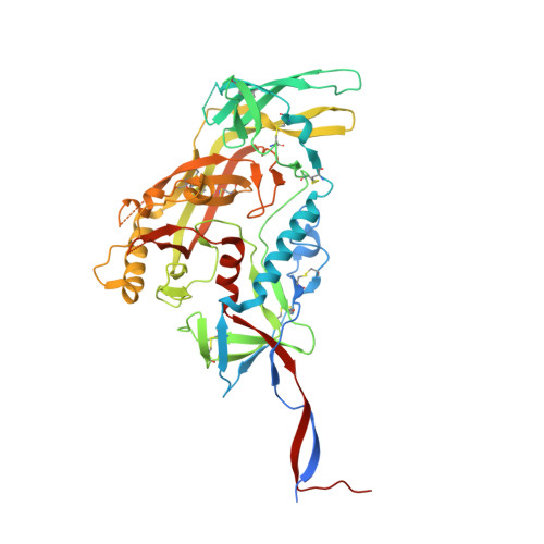
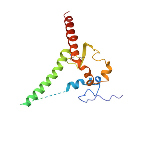
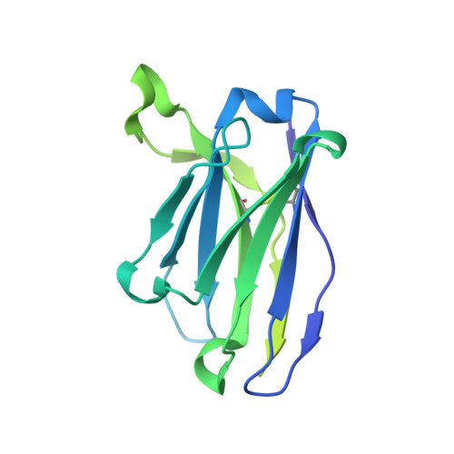
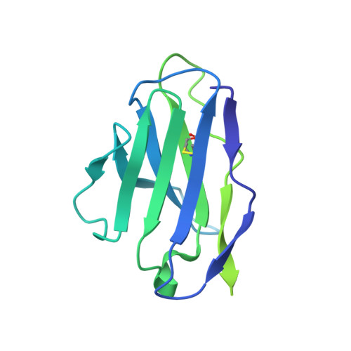
    The fusion peptide (FP) of HIV-1 envelope glycoprotein (Env) is essential for mediating viral entry. Detection of broadly neutralizing antibodies (bnAbs) that interact with the FP has revealed it as a site of vulnerability. We delineate X-ray and cryo-electron microscopy (cryo-EM) structures of bnAb ACS202, from an HIV-infected elite neutralizer, with an FP and with a soluble Env trimer (AMC011 SOSIP.v4.2) derived from the same patient. We show that ACS202 CDRH3 forms a "β strand" interaction with the exposed hydrophobic FP and recognizes a continuous region of gp120, including a conserved N-linked glycan at N88. A cryo-EM structure of another previously identified bnAb VRC34.01 with AMC011 SOSIP.v4.2 shows that it also penetrates through glycans to target the FP. We further demonstrate that the FP can twist and present different conformations for recognition by bnAbs, which enables approach to Env from diverse angles. The variable recognition of FP by bnAbs thus provides insights for vaccine design.


  • Organizational Affiliation

    Department of Integrative Structural and Computational Biology, The Scripps Research Institute, La Jolla, CA 92037, USA.


Macromolecules
Find similar proteins by:  (by identity cutoff)  |  3D Structure
Entity ID: 1
MoleculeChains Sequence LengthOrganismDetailsImage
HIV-1 Env AMC011 v4.2 SOSIP gp120512Human immunodeficiency virus 1Mutation(s): 0 
Entity Groups  
Sequence Clusters30% Identity50% Identity70% Identity90% Identity95% Identity100% Identity
Sequence Annotations
Expand
  • Reference Sequence
Find similar proteins by:  (by identity cutoff)  |  3D Structure
Entity ID: 2
MoleculeChains Sequence LengthOrganismDetailsImage
HIV-1 Env AMC011 v4.2 SOSIP gp41153Human immunodeficiency virus 1Mutation(s): 0 
UniProt
Find proteins for Q78156 (Human immunodeficiency virus 1)
Explore Q78156 
Go to UniProtKB:  Q78156
Entity Groups  
Sequence Clusters30% Identity50% Identity70% Identity90% Identity95% Identity100% Identity
UniProt GroupQ78156
Sequence Annotations
Expand
  • Reference Sequence
Find similar proteins by:  (by identity cutoff)  |  3D Structure
Entity ID: 3
MoleculeChains Sequence LengthOrganismDetailsImage
Monoclonal antibody VRC34.01 fragment antigen binding heavy chain222Homo sapiensMutation(s): 0 
Entity Groups  
Sequence Clusters30% Identity50% Identity70% Identity90% Identity95% Identity100% Identity
Sequence Annotations
Expand
  • Reference Sequence
Find similar proteins by:  (by identity cutoff)  |  3D Structure
Entity ID: 4
MoleculeChains Sequence LengthOrganismDetailsImage
Monoclonal antibody VRC34.01 fragment antigen binding light chain210Homo sapiensMutation(s): 0 
Entity Groups  
Sequence Clusters30% Identity50% Identity70% Identity90% Identity95% Identity100% Identity
Sequence Annotations
Expand
  • Reference Sequence
Oligosaccharides

Help

Entity ID: 5
MoleculeChains Length2D Diagram Glycosylation3D Interactions
alpha-D-mannopyranose-(1-3)-[alpha-D-mannopyranose-(1-6)]beta-D-mannopyranose-(1-4)-2-acetamido-2-deoxy-beta-D-glucopyranose-(1-4)-2-acetamido-2-deoxy-beta-D-glucopyranose
AB [auth 0],
BA [auth b],
DB [auth 3],
FA [auth f],
HB [auth 7],
AB [auth 0],
BA [auth b],
DB [auth 3],
FA [auth f],
HB [auth 7],
IA [auth i],
KB [auth AA],
MA [auth m],
PA [auth p],
TA [auth t],
WA [auth w],
Y
5N-Glycosylation
Glycosylation Resources
GlyTouCan:  G22768VO
GlyCosmos:  G22768VO
GlyGen:  G22768VO
Entity ID: 6
MoleculeChains Length2D Diagram Glycosylation3D Interactions
2-acetamido-2-deoxy-beta-D-glucopyranose-(1-4)-2-acetamido-2-deoxy-beta-D-glucopyranose
BB [auth 1],
CA [auth c],
EB [auth 4],
GA [auth g],
IB [auth 8],
BB [auth 1],
CA [auth c],
EB [auth 4],
GA [auth g],
IB [auth 8],
JA [auth j],
LB [auth BA],
NA [auth n],
QA [auth q],
UA [auth u],
XA [auth x],
Z
2N-Glycosylation
Glycosylation Resources
GlyTouCan:  G42666HT
GlyCosmos:  G42666HT
GlyGen:  G42666HT
Entity ID: 7
MoleculeChains Length2D Diagram Glycosylation3D Interactions
alpha-D-mannopyranose-(1-6)-beta-D-mannopyranose-(1-4)-2-acetamido-2-deoxy-beta-D-glucopyranose-(1-4)-2-acetamido-2-deoxy-beta-D-glucopyranose
AA [auth a],
CB [auth 2],
HA [auth h],
JB [auth 9],
OA [auth o],
AA [auth a],
CB [auth 2],
HA [auth h],
JB [auth 9],
OA [auth o],
VA [auth v]
4N-Glycosylation
Glycosylation Resources
GlyTouCan:  G22573RC
GlyCosmos:  G22573RC
GlyGen:  G22573RC
Entity ID: 8
MoleculeChains Length2D Diagram Glycosylation3D Interactions
beta-D-mannopyranose-(1-4)-2-acetamido-2-deoxy-beta-D-glucopyranose-(1-4)-2-acetamido-2-deoxy-beta-D-glucopyranose
DA [auth d],
EA [auth e],
FB [auth 5],
GB [auth 6],
KA [auth k],
DA [auth d],
EA [auth e],
FB [auth 5],
GB [auth 6],
KA [auth k],
LA [auth l],
MB [auth CA],
NB [auth DA],
RA [auth r],
SA [auth s],
YA [auth y],
ZA [auth z]
3N-Glycosylation
Glycosylation Resources
GlyTouCan:  G15407YE
GlyCosmos:  G15407YE
GlyGen:  G15407YE
Small Molecules
Ligands 1 Unique
IDChains Name / Formula / InChI Key2D Diagram3D Interactions
NAG
Query on NAG

Download Ideal Coordinates CCD File 
AC [auth F]
BC [auth F]
CC [auth F]
DC [auth G]
EC [auth G]
AC [auth F],
BC [auth F],
CC [auth F],
DC [auth G],
EC [auth G],
FC [auth G],
OB [auth A],
PB [auth A],
QB [auth A],
RB [auth C],
SB [auth C],
TB [auth C],
UB [auth D],
VB [auth D],
WB [auth D],
XB [auth E],
YB [auth E],
ZB [auth E]
2-acetamido-2-deoxy-beta-D-glucopyranose
C8 H15 N O6
OVRNDRQMDRJTHS-FMDGEEDCSA-N
Experimental Data & Validation

Experimental Data

  • Method: ELECTRON MICROSCOPY
  • Resolution: 4.50 Å
  • Aggregation State: PARTICLE 
  • Reconstruction Method: SINGLE PARTICLE 
EM Software:
TaskSoftware PackageVersion
MODEL REFINEMENTRosetta3.10
RECONSTRUCTIONcryoSPARC

Structure Validation

View Full Validation Report


Entry History & Funding Information

Deposition Data


Funding OrganizationLocationGrant Number
National Institutes of Health/National Institute Of Allergy and Infectious Diseases (NIH/NIAID)United StatesP01 AI110657
Bill & Melinda Gates FoundationUnited StatesOPP1115782
Bill & Melinda Gates FoundationUnited StatesOPP1084519
National Institutes of Health/National Institute Of Allergy and Infectious Diseases (NIH/NIAID)United StatesF31 Al131873
Other privateUnited States109718-63-RKNT

Revision History  (Full details and data files)

  • Version 1.0: 2019-06-19
    Type: Initial release
  • Version 1.1: 2019-06-26
    Changes: Data collection, Database references
  • Version 1.2: 2019-12-18
    Changes: Author supporting evidence
  • Version 2.0: 2020-07-29
    Type: Remediation
    Reason: Carbohydrate remediation
    Changes: Advisory, Atomic model, Data collection, Derived calculations, Other, Structure summary