5ODZ

CRYSTAL STRUCTURE OF THE BETA-LACTAMASE OXA-163


Experimental Data Snapshot

  • Method: X-RAY DIFFRACTION
  • Resolution: 2.07 Å
  • R-Value Free: 0.187 
  • R-Value Work: 0.145 
  • R-Value Observed: 0.147 

wwPDB Validation   3D Report Full Report


This is version 1.2 of the entry. See complete history


Literature

Structure, activity and thermostability investigations of OXA-163, OXA-181 and OXA-245 using biochemical analysis, crystal structures and differential scanning calorimetry analysis.

Lund, B.A.Thomassen, A.M.Carlsen, T.J.O.Leiros, H.K.S.

(2017) Acta Crystallogr F Struct Biol Commun 73: 579-587

  • DOI: https://doi.org/10.1107/S2053230X17013838
  • Primary Citation of Related Structures:  
    5ODZ, 5OE0, 5OE2

  • PubMed Abstract: 

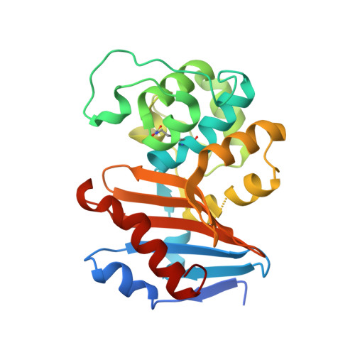
    The first crystal structures of the class D β-lactamases OXA-181 and OXA-245 were determined to 2.05 and 2.20 Å resolution, respectively; in addition, the structure of a new crystal form of OXA-163 was resolved to 2.07 Å resolution. All of these enzymes are OXA-48-like and have been isolated from different clinical Klebsiella pneumoniae strains and also from other human pathogens such as Pseudomonas aeruginosa and Escherichia coli. Here, enzyme kinetics and thermostability studies are presented, and the new crystal structures are used to explain the observed variations. OXA-245 had the highest melting point (T m = 55.8°C), as determined by differential scanning calorimetry, compared with OXA-163 (T m = 49.4°C) and OXA-181 (T m = 52.6°C). The differences could be explained by the loss of two salt bridges in OXA-163, and an overall decrease in the polarity of the surface of OXA-181 compared with OXA-245.


  • Organizational Affiliation

    Department of Chemistry, UiT The Arctic University of Norway, 9037 Tromsø, Norway.


Macromolecules
Find similar proteins by:  (by identity cutoff)  |  3D Structure
Entity ID: 1
MoleculeChains Sequence LengthOrganismDetailsImage
Beta-lactamaseA [auth B],
B [auth D]
252Enterobacter cloacaeMutation(s): 0 
Gene Names: blaOXA-163
EC: 3.5.2.6
UniProt
Find proteins for F6KZJ2 (Enterobacter cloacae)
Explore F6KZJ2 
Go to UniProtKB:  F6KZJ2
Entity Groups  
Sequence Clusters30% Identity50% Identity70% Identity90% Identity95% Identity100% Identity
UniProt GroupF6KZJ2
Sequence Annotations
Expand
  • Reference Sequence
Small Molecules
Modified Residues  1 Unique
IDChains TypeFormula2D DiagramParent
KCX
Query on KCX
A [auth B],
B [auth D]
L-PEPTIDE LINKINGC7 H14 N2 O4LYS
Experimental Data & Validation

Experimental Data

  • Method: X-RAY DIFFRACTION
  • Resolution: 2.07 Å
  • R-Value Free: 0.187 
  • R-Value Work: 0.145 
  • R-Value Observed: 0.147 
  • Space Group: P 65 2 2
Unit Cell:
Length ( Å )Angle ( ˚ )
a = 121.92α = 90
b = 121.92β = 90
c = 160.426γ = 120
Software Package:
Software NamePurpose
PHENIXrefinement
XDSdata reduction
Aimlessdata scaling
PHASERphasing

Structure Validation

View Full Validation Report


Entry History 

Deposition Data

Revision History  (Full details and data files)

  • Version 1.0: 2017-10-18
    Type: Initial release
  • Version 1.1: 2017-12-06
    Changes: Database references
  • Version 1.2: 2024-01-17
    Changes: Data collection, Database references, Derived calculations, Refinement description