5KY2

mouse POFUT1 in complex with O-glucosylated mouse Factor VII EGF1 and GDP


Experimental Data Snapshot

  • Method: X-RAY DIFFRACTION
  • Resolution: 1.47 Å
  • R-Value Free: 0.175 
  • R-Value Work: 0.153 
  • R-Value Observed: 0.154 

wwPDB Validation   3D Report Full Report


Ligand Structure Quality Assessment 


This is version 2.0 of the entry. See complete history


Literature

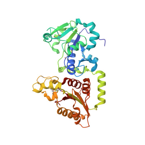
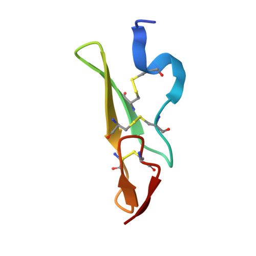
Recognition of EGF-like domains by the Notch-modifying O-fucosyltransferase POFUT1.

Li, Z.Han, K.Pak, J.E.Satkunarajah, M.Zhou, D.Rini, J.M.

(2017) Nat Chem Biol 13: 757-763

  • DOI: https://doi.org/10.1038/nchembio.2381
  • Primary Citation of Related Structures:  
    5KXH, 5KXQ, 5KY0, 5KY2, 5KY3, 5KY4, 5KY5, 5KY7, 5KY8, 5KY9

  • PubMed Abstract: 

    Protein O-fucosyltransferase 1 (POFUT1) fucosylates the epidermal growth factor (EGF)-like domains found in cell-surface and secreted glycoproteins including Notch and its ligands. Although Notch fucosylation is critical for development, and POFUT1 deficiency leads to human disease, how this enzyme binds and catalyzes the fucosylation of its diverse EGF-like domain substrates has not been determined. Reported here is the X-ray crystal structure of mouse POFUT1 in complex with several EGF-like domains, including EGF12 and EGF26 of Notch. Overall shape complementarity, interactions with invariant atoms of the fucosylation motif and flexible segments on POFUT1 all define its EGF-like-domain binding properties. Using large-scale structural and sequence analysis, we also show that POFUT1 binds EGF-like domains of the hEGF type and that the highly correlated presence of POFUT1 and fucosylatable hEGFs has accompanied animal evolution.


  • Organizational Affiliation

    Department of Biochemistry, University of Toronto, Toronto, Ontario, Canada.


Macromolecules
Find similar proteins by:  (by identity cutoff)  |  3D Structure
Entity ID: 1
MoleculeChains Sequence LengthOrganismDetailsImage
GDP-fucose protein O-fucosyltransferase 1355Mus musculusMutation(s): 0 
Gene Names: Pofut1
EC: 2.4.1.221
UniProt
Find proteins for Q91ZW2 (Mus musculus)
Explore Q91ZW2 
Go to UniProtKB:  Q91ZW2
Entity Groups  
Sequence Clusters30% Identity50% Identity70% Identity90% Identity95% Identity100% Identity
UniProt GroupQ91ZW2
Sequence Annotations
Expand
  • Reference Sequence
Find similar proteins by:  (by identity cutoff)  |  3D Structure
Entity ID: 2
MoleculeChains Sequence LengthOrganismDetailsImage
Coagulation factor VII40Mus musculusMutation(s): 0 
Gene Names: F7Cf7
EC: 3.4.21.21
UniProt & NIH Common Fund Data Resources
Find proteins for P70375 (Mus musculus)
Explore P70375 
Go to UniProtKB:  P70375
IMPC:  MGI:109325
Entity Groups  
Sequence Clusters30% Identity50% Identity70% Identity90% Identity95% Identity100% Identity
UniProt GroupP70375
Sequence Annotations
Expand
  • Reference Sequence
Small Molecules
Ligands 4 Unique
IDChains Name / Formula / InChI Key2D Diagram3D Interactions
GDP
Query on GDP

Download Ideal Coordinates CCD File 
E [auth A]GUANOSINE-5'-DIPHOSPHATE
C10 H15 N5 O11 P2
QGWNDRXFNXRZMB-UUOKFMHZSA-N
NAG
Query on NAG

Download Ideal Coordinates CCD File 
C [auth A],
D [auth A]
2-acetamido-2-deoxy-beta-D-glucopyranose
C8 H15 N O6
OVRNDRQMDRJTHS-FMDGEEDCSA-N
BGC
Query on BGC

Download Ideal Coordinates CCD File 
I [auth B]beta-D-glucopyranose
C6 H12 O6
WQZGKKKJIJFFOK-VFUOTHLCSA-N
GOL
Query on GOL

Download Ideal Coordinates CCD File 
F [auth A],
G [auth A],
H [auth A]
GLYCEROL
C3 H8 O3
PEDCQBHIVMGVHV-UHFFFAOYSA-N
Experimental Data & Validation

Experimental Data

  • Method: X-RAY DIFFRACTION
  • Resolution: 1.47 Å
  • R-Value Free: 0.175 
  • R-Value Work: 0.153 
  • R-Value Observed: 0.154 
  • Space Group: P 21 21 21
Unit Cell:
Length ( Å )Angle ( ˚ )
a = 51.965α = 90
b = 67.138β = 90
c = 109.357γ = 90
Software Package:
Software NamePurpose
XSCALEdata scaling
PHENIXrefinement
PDB_EXTRACTdata extraction
XDSdata reduction
PHASERphasing

Structure Validation

View Full Validation Report


Ligand Structure Quality Assessment 


Entry History & Funding Information

Deposition Data


Funding OrganizationLocationGrant Number
Canadian Institutes of Health Research (CIHR)Canada--

Revision History  (Full details and data files)

  • Version 1.0: 2017-05-17
    Type: Initial release
  • Version 1.1: 2017-05-31
    Changes: Database references
  • Version 1.2: 2017-06-28
    Changes: Database references
  • Version 1.3: 2017-09-20
    Changes: Author supporting evidence
  • Version 1.4: 2020-01-08
    Changes: Author supporting evidence, Data collection
  • Version 2.0: 2020-07-29
    Type: Remediation
    Reason: Carbohydrate remediation
    Changes: Atomic model, Data collection, Derived calculations, Structure summary