5CX7

Crystal Structure of PduOC:Heme Complex


Experimental Data Snapshot

  • Method: X-RAY DIFFRACTION
  • Resolution: 1.97 Å
  • R-Value Free: 0.186 
  • R-Value Work: 0.158 
  • R-Value Observed: 0.159 

wwPDB Validation   3D Report Full Report


Ligand Structure Quality Assessment 


This is version 1.3 of the entry. See complete history


Literature

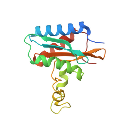
The Crystal Structure of the C-Terminal Domain of the Salmonella enterica PduO Protein: An Old Fold with a New Heme-Binding Mode.

Ortiz de Orue Lucana, D.Hickey, N.Hensel, M.Klare, J.P.Geremia, S.Tiufiakova, T.Torda, A.E.

(2016) Front Microbiol 7: 1010-1010

  • DOI: https://doi.org/10.3389/fmicb.2016.01010
  • Primary Citation of Related Structures:  
    5CX7

  • PubMed Abstract: 

    The two-domain protein PduO, involved in 1,2-propanediol utilization in the pathogenic Gram-negative bacterium Salmonella enterica is an ATP:Cob(I)alamin adenosyltransferase, but this is a function of the N-terminal domain alone. The role of its C-terminal domain (PduOC) is, however, unknown. In this study, comparative growth assays with a set of Salmonella mutant strains showed that this domain is necessary for effective in vivo catabolism of 1,2-propanediol. It was also shown that isolated, recombinantly-expressed PduOC binds heme in vivo. The structure of PduOC co-crystallized with heme was solved (1.9 Å resolution) showing an octameric assembly with four heme moieities. The four heme groups are highly solvent-exposed and the heme iron is hexa-coordinated with bis-His ligation by histidines from different monomers. Static light scattering confirmed the octameric assembly in solution, but a mutation of the heme-coordinating histidine caused dissociation into dimers. Isothermal titration calorimetry using the PduOC apoprotein showed strong heme binding (K d = 1.6 × 10(-7) M). Biochemical experiments showed that the absence of the C-terminal domain in PduO did not affect adenosyltransferase activity in vitro. The evidence suggests that PduOC:heme plays an important role in the set of cobalamin transformations required for effective catabolism of 1,2-propanediol. Salmonella PduO is one of the rare proteins which binds the redox-active metabolites heme and cobalamin, and the heme-binding mode of the C-terminal domain differs from that in other members of this protein family.


  • Organizational Affiliation

    Applied Genetics of Microorganisms, Department of Biology/Chemistry, University of Osnabrück Osnabrück, Germany.


Macromolecules
Find similar proteins by:  (by identity cutoff)  |  3D Structure
Entity ID: 1
MoleculeChains Sequence LengthOrganismDetailsImage
ATP:cob(I)alamin adenosyltransferase
A, B, C, D, E
A, B, C, D, E, F, G, H, I, J, K, L, M, N, O, P
147Salmonella enterica subsp. enterica serovar LivingstoneMutation(s): 0 
Gene Names: VN13_17650
UniProt
Find proteins for Q8ZNR5 (Salmonella typhimurium (strain LT2 / SGSC1412 / ATCC 700720))
Explore Q8ZNR5 
Go to UniProtKB:  Q8ZNR5
Entity Groups  
Sequence Clusters30% Identity50% Identity70% Identity90% Identity95% Identity100% Identity
UniProt GroupQ8ZNR5
Sequence Annotations
Expand
  • Reference Sequence
Small Molecules
Ligands 5 Unique
IDChains Name / Formula / InChI Key2D Diagram3D Interactions
HEM
Query on HEM

Download Ideal Coordinates CCD File 
FA [auth E]
FB [auth K]
KA [auth G]
KB [auth M]
Q [auth A]
FA [auth E],
FB [auth K],
KA [auth G],
KB [auth M],
Q [auth A],
QB [auth O],
TA [auth I],
Y [auth C]
PROTOPORPHYRIN IX CONTAINING FE
C34 H32 Fe N4 O4
KABFMIBPWCXCRK-RGGAHWMASA-L
GOL
Query on GOL

Download Ideal Coordinates CCD File 
BA [auth D]
BB [auth J]
CA [auth D]
GA [auth E]
GB [auth K]
BA [auth D],
BB [auth J],
CA [auth D],
GA [auth E],
GB [auth K],
HB [auth K],
IA [auth F],
IB [auth K],
LA [auth G],
LB [auth M],
MA [auth G],
NA [auth G],
OB [auth N],
PA [auth H],
R [auth A],
RB [auth O],
S [auth A],
T [auth A],
TB [auth P],
UA [auth I],
VA [auth I],
W [auth B],
WA [auth I],
XA [auth I],
Z [auth C]
GLYCEROL
C3 H8 O3
PEDCQBHIVMGVHV-UHFFFAOYSA-N
CL
Query on CL

Download Ideal Coordinates CCD File 
AA [auth C],
DB [auth J],
EA [auth D],
ZA [auth I]
CHLORIDE ION
Cl
VEXZGXHMUGYJMC-UHFFFAOYSA-M
MG
Query on MG

Download Ideal Coordinates CCD File 
QA [auth H],
UB [auth P]
MAGNESIUM ION
Mg
JLVVSXFLKOJNIY-UHFFFAOYSA-N
NA
Query on NA

Download Ideal Coordinates CCD File 
AB [auth I]
CB [auth J]
DA [auth D]
EB [auth J]
HA [auth E]
AB [auth I],
CB [auth J],
DA [auth D],
EB [auth J],
HA [auth E],
JA [auth F],
JB [auth L],
MB [auth M],
NB [auth M],
OA [auth G],
PB [auth N],
RA [auth H],
SA [auth H],
SB [auth P],
U [auth A],
V [auth A],
X [auth B],
YA [auth I]
SODIUM ION
Na
FKNQFGJONOIPTF-UHFFFAOYSA-N
Experimental Data & Validation

Experimental Data

  • Method: X-RAY DIFFRACTION
  • Resolution: 1.97 Å
  • R-Value Free: 0.186 
  • R-Value Work: 0.158 
  • R-Value Observed: 0.159 
  • Space Group: P 1 21 1
Unit Cell:
Length ( Å )Angle ( ˚ )
a = 71.3α = 90
b = 130.12β = 90
c = 120.75γ = 90
Software Package:
Software NamePurpose
REFMACrefinement
MOSFLMdata reduction
SCALAdata scaling
AMoREphasing

Structure Validation

View Full Validation Report


Ligand Structure Quality Assessment 


Entry History 

Deposition Data

Revision History  (Full details and data files)

  • Version 1.0: 2016-06-29
    Type: Initial release
  • Version 1.1: 2016-08-03
    Changes: Database references
  • Version 1.2: 2016-11-23
    Changes: Derived calculations
  • Version 1.3: 2024-01-10
    Changes: Data collection, Database references, Derived calculations, Refinement description