5FKP

Crystal structure of the mouse CD1d in complex with the p99 peptide


Experimental Data Snapshot

  • Method: X-RAY DIFFRACTION
  • Resolution: 1.80 Å
  • R-Value Free: 0.234 
  • R-Value Work: 0.205 
  • R-Value Observed: 0.207 

wwPDB Validation   3D Report Full Report


Ligand Structure Quality Assessment 


This is version 2.1 of the entry. See complete history


Literature

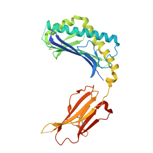
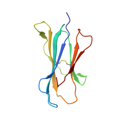
Structure of an Alpha-Helical Peptide and Lipopeptide Bound to the Non-Classical Mhc Class I Molecule Cd1D

Girardi, E.Wang, J.Zajonc, D.M.

(2016) J Biol Chem 291: 10677

  • DOI: https://doi.org/10.1074/jbc.M115.702118
  • Primary Citation of Related Structures:  
    5EFI, 5FKP

  • PubMed Abstract: 

    Mouse CD1d is a nonclassical MHC molecule able to present lipids and glycolipids to a specialized subset of T cells known as natural killer T cells. The antigens presented by CD1d have been shown to cover a broad range of chemical structures and to follow precise rules determining the potency of the antigen in the context of T cell activation. Together with lipids, initial reports suggested that CD1d can also bind and present hydrophobic peptides with (F/W)XX(I/L/M)XXW. However, the exact location of peptide binding and the molecular basis for the required motif are currently unknown. Here we present the crystal structure of the first peptide identified to bind CD1d, p99, and show that it binds in the antigen-binding groove of CD1d in a manner compatible with its presentation to T cell receptors. Interestingly, the peptide adopts an α-helical conformation, which orients the motif residues toward its deep binding groove, therefore explaining the molecular requirements for peptide binding. Moreover, we demonstrate that a lipopeptide version of the same peptide is able to bind CD1d in a similar conformation, identifying another class of molecules binding this antigen-presenting molecule.


  • Organizational Affiliation

    From the Division of Cell Biology, La Jolla Institute for Allergy and Immunology, La Jolla, California 92037 and.


Macromolecules
Find similar proteins by:  (by identity cutoff)  |  3D Structure
Entity ID: 1
MoleculeChains Sequence LengthOrganismDetailsImage
ANTIGEN-PRESENTING GLYCOPROTEIN CD1D1285Mus musculusMutation(s): 0 
UniProt & NIH Common Fund Data Resources
Find proteins for P11609 (Mus musculus)
Explore P11609 
Go to UniProtKB:  P11609
IMPC:  MGI:107674
Entity Groups  
Sequence Clusters30% Identity50% Identity70% Identity90% Identity95% Identity100% Identity
UniProt GroupP11609
Sequence Annotations
Expand
  • Reference Sequence
Find similar proteins by:  (by identity cutoff)  |  3D Structure
Entity ID: 2
MoleculeChains Sequence LengthOrganismDetailsImage
BETA 2 MICROGLOBULIN99Mus musculusMutation(s): 0 
UniProt & NIH Common Fund Data Resources
Find proteins for P01887 (Mus musculus)
Explore P01887 
Go to UniProtKB:  P01887
IMPC:  MGI:88127
Entity Groups  
Sequence Clusters30% Identity50% Identity70% Identity90% Identity95% Identity100% Identity
UniProt GroupP01887
Sequence Annotations
Expand
  • Reference Sequence

Find similar proteins by:  Sequence   |   3D Structure  

Entity ID: 3
MoleculeChains Sequence LengthOrganismDetailsImage
P9922Mus musculusMutation(s): 0 
Entity Groups  
Sequence Clusters30% Identity50% Identity70% Identity90% Identity95% Identity100% Identity
Sequence Annotations
Expand
  • Reference Sequence
Oligosaccharides

Help

Entity ID: 4
MoleculeChains Length2D Diagram Glycosylation3D Interactions
alpha-D-mannopyranose-(1-6)-beta-D-mannopyranose-(1-4)-2-acetamido-2-deoxy-beta-D-glucopyranose-(1-4)-[alpha-L-fucopyranose-(1-6)]2-acetamido-2-deoxy-beta-D-glucopyranose
D
5N-Glycosylation
Glycosylation Resources
GlyTouCan:  G00395TQ
GlyCosmos:  G00395TQ
GlyGen:  G00395TQ
Small Molecules
Ligands 4 Unique
IDChains Name / Formula / InChI Key2D Diagram3D Interactions
6UL
Query on 6UL

Download Ideal Coordinates CCD File 
G [auth A]TETRACOSYL PALMITATE
C40 H80 O2
KKTAIMZGSAQXRE-UHFFFAOYSA-N
NAG
Query on NAG

Download Ideal Coordinates CCD File 
E [auth A],
F [auth A]
2-acetamido-2-deoxy-beta-D-glucopyranose
C8 H15 N O6
OVRNDRQMDRJTHS-FMDGEEDCSA-N
TAR
Query on TAR

Download Ideal Coordinates CCD File 
L [auth B]D(-)-TARTARIC ACID
C4 H6 O6
FEWJPZIEWOKRBE-LWMBPPNESA-N
GOL
Query on GOL

Download Ideal Coordinates CCD File 
H [auth A],
I [auth A],
J [auth A],
K [auth B]
GLYCEROL
C3 H8 O3
PEDCQBHIVMGVHV-UHFFFAOYSA-N
Experimental Data & Validation

Experimental Data

  • Method: X-RAY DIFFRACTION
  • Resolution: 1.80 Å
  • R-Value Free: 0.234 
  • R-Value Work: 0.205 
  • R-Value Observed: 0.207 
  • Space Group: P 21 21 21
Unit Cell:
Length ( Å )Angle ( ˚ )
a = 42.17α = 90
b = 107.57β = 90
c = 110.61γ = 90
Software Package:
Software NamePurpose
REFMACrefinement
MOSFLMdata reduction
SCALAdata scaling
MOLREPphasing

Structure Validation

View Full Validation Report


Ligand Structure Quality Assessment 


Entry History 

Deposition Data

Revision History  (Full details and data files)

  • Version 1.0: 2016-03-30
    Type: Initial release
  • Version 1.1: 2016-04-06
    Changes: Database references
  • Version 1.2: 2016-06-01
    Changes: Database references
  • Version 2.0: 2020-07-29
    Type: Remediation
    Reason: Carbohydrate remediation
    Changes: Advisory, Atomic model, Data collection, Derived calculations, Other, Structure summary
  • Version 2.1: 2024-01-10
    Changes: Data collection, Database references, Refinement description, Structure summary