5E0Q

Crystal structure of the Nup98 C-terminal domain bound to nanobody TP377


Experimental Data Snapshot

  • Method: X-RAY DIFFRACTION
  • Resolution: 1.90 Å
  • R-Value Free: 0.196 
  • R-Value Work: 0.167 
  • R-Value Observed: 0.169 

wwPDB Validation   3D Report Full Report


This is version 1.2 of the entry. See complete history

Re-refinement Note

A newer entry is available that reflects an alternative modeling of the original data: 7NOW


Literature

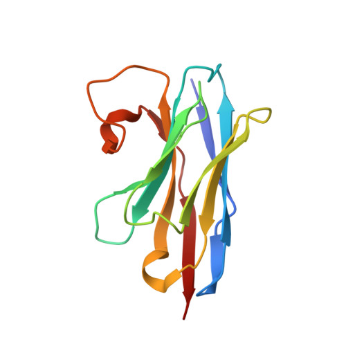
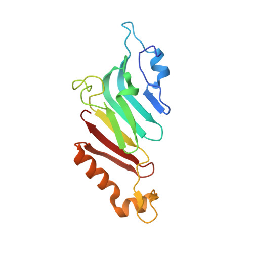
Nanobodies: site-specific labeling for super-resolution imaging, rapid epitope-mapping and native protein complex isolation.

Pleiner, T.Bates, M.Trakhanov, S.Lee, C.T.Schliep, J.E.Chug, H.Bohning, M.Stark, H.Urlaub, H.Gorlich, D.

(2015) Elife 4: e11349-e11349

  • DOI: https://doi.org/10.7554/eLife.11349
  • Primary Citation of Related Structures:  
    5E0Q

  • PubMed Abstract: 

    Nanobodies are single-domain antibodies of camelid origin. We generated nanobodies against the vertebrate nuclear pore complex (NPC) and used them in STORM imaging to locate individual NPC proteins with <2 nm epitope-label displacement. For this, we introduced cysteines at specific positions in the nanobody sequence and labeled the resulting proteins with fluorophore-maleimides. As nanobodies are normally stabilized by disulfide-bonded cysteines, this appears counterintuitive. Yet, our analysis showed that this caused no folding problems. Compared to traditional NHS ester-labeling of lysines, the cysteine-maleimide strategy resulted in far less background in fluorescence imaging, it better preserved epitope recognition and it is site-specific. We also devised a rapid epitope-mapping strategy, which relies on crosslinking mass spectrometry and the introduced ectopic cysteines. Finally, we used different anti-nucleoporin nanobodies to purify the major NPC building blocks – each in a single step, with native elution and, as demonstrated, in excellent quality for structural analysis by electron microscopy. The presented strategies are applicable to any nanobody and nanobody-target.


  • Organizational Affiliation

    Department of Cellular Logistics, Max Planck Institute for Biophysical Chemistry, Göttingen, Germany.


Macromolecules
Find similar proteins by:  (by identity cutoff)  |  3D Structure
Entity ID: 1
MoleculeChains Sequence LengthOrganismDetailsImage
Anti-Nup98 Nanobody TP377128Vicugna pacosMutation(s): 0 
Entity Groups  
Sequence Clusters30% Identity50% Identity70% Identity90% Identity95% Identity100% Identity
Sequence Annotations
Expand
  • Reference Sequence
Find similar proteins by:  (by identity cutoff)  |  3D Structure
Entity ID: 2
MoleculeChains Sequence LengthOrganismDetailsImage
Nuclear pore complex protein Nup98-Nup96153Xenopus tropicalisMutation(s): 1 
Gene Names: nup98
UniProt
Find proteins for J7I6Y1 (Xenopus tropicalis)
Explore J7I6Y1 
Go to UniProtKB:  J7I6Y1
Entity Groups  
Sequence Clusters30% Identity50% Identity70% Identity90% Identity95% Identity100% Identity
UniProt GroupJ7I6Y1
Sequence Annotations
Expand
  • Reference Sequence
Experimental Data & Validation

Experimental Data

  • Method: X-RAY DIFFRACTION
  • Resolution: 1.90 Å
  • R-Value Free: 0.196 
  • R-Value Work: 0.167 
  • R-Value Observed: 0.169 
  • Space Group: P 41
Unit Cell:
Length ( Å )Angle ( ˚ )
a = 66.59α = 90
b = 66.59β = 90
c = 87.9γ = 90
Software Package:
Software NamePurpose
PHENIXrefinement
XDSdata reduction
XSCALEdata scaling
PHENIXphasing

Structure Validation

View Full Validation Report


Entry History & Funding Information

Deposition Data


Funding OrganizationLocationGrant Number
DeutscheGermany--

Revision History  (Full details and data files)

  • Version 1.0: 2015-12-16
    Type: Initial release
  • Version 1.1: 2016-04-27
    Changes: Database references
  • Version 1.2: 2024-01-10
    Changes: Data collection, Database references, Refinement description