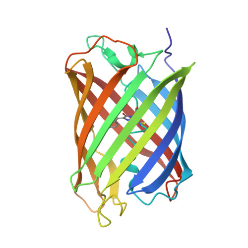
Low-Temperature Chromophore Isomerization Reveals the Photoswitching Mechanism of the Fluorescent Protein Padron.
Regis Faro, A., Carpentier, P., Jonasson, G., Pompidor, G., Arcizet, D., Demachy, I., Bourgeois, D.(2011) J Am Chem Soc 133: 16362
- PubMed: 21923132
- DOI: https://doi.org/10.1021/ja207001y
- Primary Citation of Related Structures:
3ZUF, 3ZUJ, 3ZUL - PubMed Abstract:
Photoactivatable fluorescent proteins are essential players in nanoscopy approaches based on the super-localization of single molecules. The subclass of reversibly photoswitchable fluorescent proteins typically activate through isomerization of the chromophore coupled with a change in its protonation state. However, the interplay between these two events, the details of photoswitching pathways, and the role of protein dynamics remain incompletely understood. Here, by using a combination of structural and spectroscopic approaches, we discovered two fluorescent intermediate states along the on-switching pathway of the fluorescent protein Padron. The first intermediate can be populated at temperatures as low as 100 K and results from a remarkable trans-cis isomerization of the anionic chromophore taking place within a protein matrix essentially deprived of conformational flexibility. This intermediate evolves in the dark at cryotemperatures to a second structurally similar but spectroscopically distinct anionic intermediate. The final fluorescent state, which consists of a mixture of anionic and neutral chromophores in the cis configuration, is only reached above the glass transition temperature, suggesting that chromophore protonation involves solvent interactions mediated by pronounced dynamical breathing of the protein scaffold. The possibility of efficiently and reversibly photoactivating Padron at cryotemperatures will facilitate the development of advanced super-resolution imaging modalities such as cryonanoscopy.
Organizational Affiliation:
Pixel Team, IBS, Institut de Biologie Structurale Jean-Pierre Ebel, CEA, CNRS, Université Joseph Fourier, 41 rue Jules Horowitz, F-38027 Grenoble, France.