







 Download Mendeley
Structural presentation of an amyloid beta fragment by HLA
Lopez-Sagaseta, J.To be published.
Experimental Data Snapshot
Starting Model: in silico
View more details
Entity ID: 1 | |||||
|---|---|---|---|---|---|
| Molecule | Chains | Sequence Length | Organism | Details | Image |
| Amyloid-beta protein 40,HLA class II histocompatibility antigen DR beta chain | A [auth B], C [auth D] | 227 | Homo sapiens | Mutation(s): 0 Gene Names: APP, A4, AD1, HLA-DRB1 | 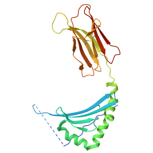 |
UniProt & NIH Common Fund Data Resources | |||||
PHAROS: P05067 GTEx: ENSG00000142192 | |||||
Entity Groups | |||||
| Sequence Clusters | 30% Identity50% Identity70% Identity90% Identity95% Identity100% Identity | ||||
| UniProt Groups | P05067D7RIG0 | ||||
Sequence AnnotationsExpand | |||||
Reference Sequence | |||||
Entity ID: 2 | |||||
|---|---|---|---|---|---|
| Molecule | Chains | Sequence Length | Organism | Details | Image |
| HLA class II histocompatibility antigen, DR alpha chain | B [auth A], D [auth C] | 198 | Homo sapiens | Mutation(s): 0 Gene Names: HLA-DRA, HLA-DRA1 | 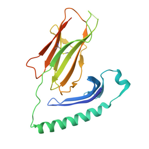 |
UniProt & NIH Common Fund Data Resources | |||||
PHAROS: P01903 GTEx: ENSG00000204287 | |||||
Entity Groups | |||||
| Sequence Clusters | 30% Identity50% Identity70% Identity90% Identity95% Identity100% Identity | ||||
| UniProt Group | P01903 | ||||
Glycosylation | |||||
| Glycosylation Sites: 1 | Go to GlyGen: P01903-1 | ||||
Sequence AnnotationsExpand | |||||
Reference Sequence | |||||
| Ligands 2 Unique | |||||
|---|---|---|---|---|---|
| ID | Chains | Name / Formula / InChI Key | 2D Diagram | 3D Interactions | |
| NAG Download:Ideal Coordinates CCD File | G [auth A] | 2-acetamido-2-deoxy-beta-D-glucopyranose C8 H15 N O6 OVRNDRQMDRJTHS-FMDGEEDCSA-N |  | ||
| EDO Download:Ideal Coordinates CCD File | E [auth A], F [auth A], H [auth C] | 1,2-ETHANEDIOL C2 H6 O2 LYCAIKOWRPUZTN-UHFFFAOYSA-N |  | ||
| Length ( Å ) | Angle ( ˚ ) |
|---|---|
| a = 57.308 | α = 90 |
| b = 118.508 | β = 109.148 |
| c = 67.126 | γ = 90 |
| Software Name | Purpose |
|---|---|
| PHENIX | refinement |
| xia2 | data reduction |
| Aimless | data scaling |
| PHASER | phasing |
| Funding Organization | Location | Grant Number |
|---|---|---|
| Other government | Spain | PC24-MEMORIAL-017-003-015 |








