2RDB

X-ray Crystal Structure of Toluene/o-Xylene Monooxygenase Hydroxylase I100W Mutant


Experimental Data Snapshot

  • Method: X-RAY DIFFRACTION
  • Resolution: 2.10 Å
  • R-Value Free: 0.284 
  • R-Value Work: 0.217 
  • R-Value Observed: 0.221 

wwPDB Validation   3D Report Full Report


Ligand Structure Quality Assessment 


This is version 1.4 of the entry. See complete history


Literature

Dioxygen activation at non-heme diiron centers: oxidation of a proximal residue in the I100W variant of toluene/o-xylene monooxygenase hydroxylase.

Murray, L.J.Garcia-Serres, R.McCormick, M.S.Davydov, R.Naik, S.G.Kim, S.H.Hoffman, B.M.Huynh, B.H.Lippard, S.J.

(2007) Biochemistry 46: 14795-14809

  • DOI: https://doi.org/10.1021/bi7017128
  • Primary Citation of Related Structures:  
    2RDB

  • PubMed Abstract: 

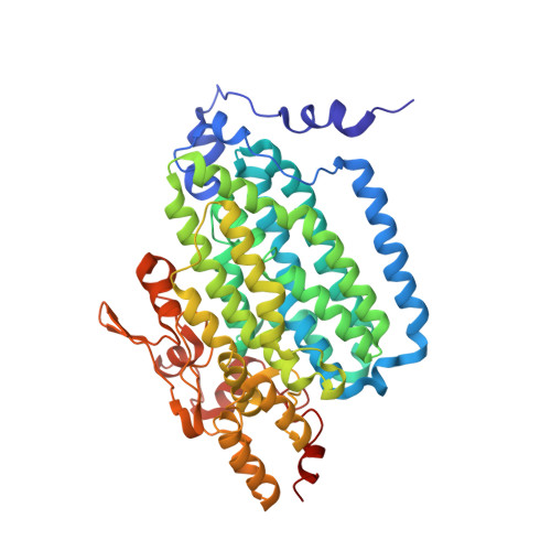
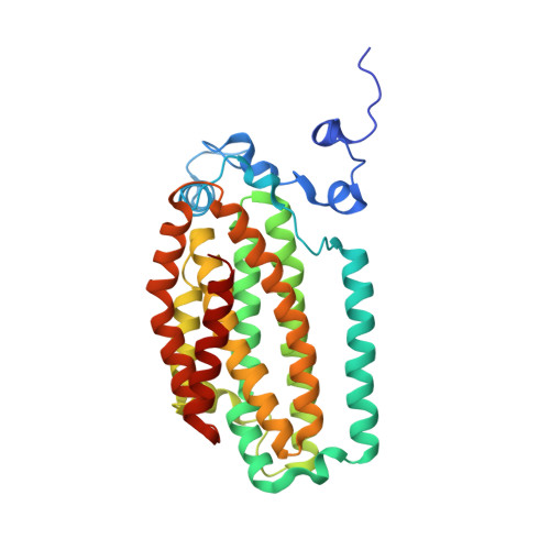
    At its carboxylate-bridged diiron active site, the hydroxylase component of toluene/o-xylene monooxygenase activates dioxygen for subsequent arene hydroxylation. In an I100W variant of this enzyme, we characterized the formation and decay of two species formed by addition of dioxygen to the reduced, diiron(II) state by rapid-freeze quench (RFQ) EPR, Mössbauer, and ENDOR spectroscopy. The dependence of the formation and decay rates of this mixed-valent transient on pH and the presence of phenol, propylene, or acetylene was investigated by double-mixing stopped-flow optical spectroscopy. Modification of the alpha-subunit of the hydroxylase after reaction of the reduced protein with dioxygen-saturated buffer was investigated by tryptic digestion coupled mass spectrometry. From these investigations, we conclude that (i) a diiron(III,IV)-W* transient, kinetically linked to a preceding diiron(III) intermediate, arises from the one-electron oxidation of W100, (ii) the tryptophan radical is deprotonated, (iii) rapid exchange of either a terminal water or hydroxide ion with water occurs at the ferric ion in the diiron(III,IV) cluster, and (iv) the diiron(III,IV) core and W* decay to the diiron(III) product by a common mechanism. No transient radical was observed by stopped-flow optical spectroscopy for reactions of the reduced hydroxylase variants I100Y, L208F, and F205W with dioxygen. The absence of such species, and the deprotonated state of the tryptophanyl radical in the diiron(III,IV)-W* transient, allow for a conservative estimate of the reduction potential of the diiron(III) intermediate as lying between 1.1 and 1.3 V. We also describe the X-ray crystal structure of the I100W variant of ToMOH.


  • Organizational Affiliation

    Department of Chemistry, Massachusetts Institute of Technology, Cambridge, Massachusetts 02139, USA.


Macromolecules
Find similar proteins by:  (by identity cutoff)  |  3D Structure
Entity ID: 1
MoleculeChains Sequence LengthOrganismDetailsImage
Toluene, o-xylene monooxygenase oxygenase subunit;alpha498Stutzerimonas stutzeriMutation(s): 1 
Gene Names: touA
UniProt
Find proteins for O87798 (Stutzerimonas stutzeri)
Explore O87798 
Go to UniProtKB:  O87798
Entity Groups  
Sequence Clusters30% Identity50% Identity70% Identity90% Identity95% Identity100% Identity
UniProt GroupO87798
Sequence Annotations
Expand
  • Reference Sequence
Find similar proteins by:  (by identity cutoff)  |  3D Structure
Entity ID: 2
MoleculeChains Sequence LengthOrganismDetailsImage
Toluene, o-xylene monooxygenase oxygenase subunit;beta330Stutzerimonas stutzeriMutation(s): 0 
Gene Names: touE
UniProt
Find proteins for O87802 (Stutzerimonas stutzeri)
Explore O87802 
Go to UniProtKB:  O87802
Entity Groups  
Sequence Clusters30% Identity50% Identity70% Identity90% Identity95% Identity100% Identity
UniProt GroupO87802
Sequence Annotations
Expand
  • Reference Sequence
Find similar proteins by:  (by identity cutoff)  |  3D Structure
Entity ID: 3
MoleculeChains Sequence LengthOrganismDetailsImage
Toluene, o-xylene monooxygenase oxygenase subunit;gamma86Stutzerimonas stutzeriMutation(s): 0 
Gene Names: touB
UniProt
Find proteins for O87799 (Stutzerimonas stutzeri)
Explore O87799 
Go to UniProtKB:  O87799
Entity Groups  
Sequence Clusters30% Identity50% Identity70% Identity90% Identity95% Identity100% Identity
UniProt GroupO87799
Sequence Annotations
Expand
  • Reference Sequence
Experimental Data & Validation

Experimental Data

  • Method: X-RAY DIFFRACTION
  • Resolution: 2.10 Å
  • R-Value Free: 0.284 
  • R-Value Work: 0.217 
  • R-Value Observed: 0.221 
  • Space Group: P 31 2 1
Unit Cell:
Length ( Å )Angle ( ˚ )
a = 182.42α = 90
b = 182.42β = 90
c = 67.962γ = 120
Software Package:
Software NamePurpose
DENZOdata reduction
SCALEPACKdata scaling
REFMACrefinement
PDB_EXTRACTdata extraction
SSRLdata collection
HKL-2000data reduction
HKL-2000data scaling
EPMRphasing

Structure Validation

View Full Validation Report


Ligand Structure Quality Assessment 


Entry History 

Deposition Data

Revision History  (Full details and data files)

  • Version 1.0: 2007-12-18
    Type: Initial release
  • Version 1.1: 2011-07-13
    Changes: Non-polymer description, Version format compliance
  • Version 1.2: 2017-10-25
    Changes: Refinement description
  • Version 1.3: 2021-10-20
    Changes: Database references, Derived calculations
  • Version 1.4: 2023-08-30
    Changes: Data collection, Refinement description