2GY7

Angiopoietin-2/Tie2 Complex Crystal Structure


Experimental Data Snapshot

  • Method: X-RAY DIFFRACTION
  • Resolution: 3.70 Å
  • R-Value Free: 0.317 
  • R-Value Work: 0.240 

wwPDB Validation   3D Report Full Report


Ligand Structure Quality Assessment 


This is version 2.0 of the entry. See complete history


Literature

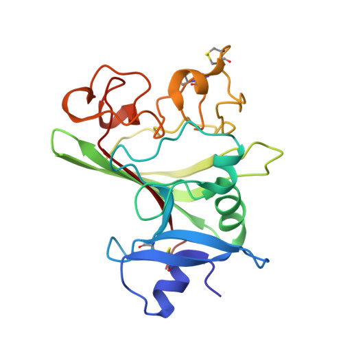
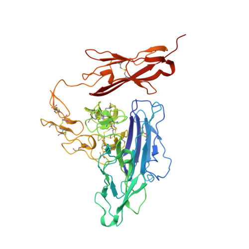
Crystal structures of the Tie2 receptor ectodomain and the angiopoietin-2-Tie2 complex.

Barton, W.A.Tzvetkova-Robev, D.Miranda, E.P.Kolev, M.V.Rajashankar, K.R.Himanen, J.P.Nikolov, D.B.

(2006) Nat Struct Mol Biol 13: 524-532

  • DOI: https://doi.org/10.1038/nsmb1101
  • Primary Citation of Related Structures:  
    2GY5, 2GY7

  • PubMed Abstract: 

    The Tie receptor tyrosine kinases and their angiopoietin (Ang) ligands play central roles in developmental and tumor-induced angiogenesis. Here we present the crystal structures of the Tie2 ligand-binding region alone and in complex with Ang2. In contrast to prediction, Tie2 contains not two but three immunoglobulin (Ig) domains, which fold together with the three epidermal growth factor domains into a compact, arrowhead-shaped structure. Ang2 binds at the tip of the arrowhead utilizing a lock-and-key mode of ligand recognition-unique for a receptor kinase-where two complementary surfaces interact with each other with no domain rearrangements and little conformational change in either molecule. Ang2-Tie2 recognition is similar to antibody-protein antigen recognition, including the location of the ligand-binding site within the Ig fold. Analysis of the structures and structure-based mutagenesis provide insight into the mechanism of receptor activation and support the hypothesis that all angiopoietins interact with Tie2 in a structurally similar manner.


  • Organizational Affiliation

    Structural Biology Program, Memorial Sloan-Kettering Cancer Center, 1275 York Avenue, New York, New York 10021, USA.


Macromolecules
Find similar proteins by:  (by identity cutoff)  |  3D Structure
Entity ID: 1
MoleculeChains Sequence LengthOrganismDetailsImage
Angiopoietin-2216Homo sapiensMutation(s): 0 
UniProt & NIH Common Fund Data Resources
Find proteins for O15123 (Homo sapiens)
Explore O15123 
Go to UniProtKB:  O15123
PHAROS:  O15123
GTEx:  ENSG00000091879 
Entity Groups  
Sequence Clusters30% Identity50% Identity70% Identity90% Identity95% Identity100% Identity
UniProt GroupO15123
Sequence Annotations
Expand
  • Reference Sequence
Find similar proteins by:  (by identity cutoff)  |  3D Structure
Entity ID: 2
MoleculeChains Sequence LengthOrganismDetailsImage
Angiopoietin-1 receptor423Homo sapiensMutation(s): 0 
Gene Names: TEKTIE2
EC: 2.7.10.1
UniProt & NIH Common Fund Data Resources
Find proteins for Q02763 (Homo sapiens)
Explore Q02763 
Go to UniProtKB:  Q02763
PHAROS:  Q02763
GTEx:  ENSG00000120156 
Entity Groups  
Sequence Clusters30% Identity50% Identity70% Identity90% Identity95% Identity100% Identity
UniProt GroupQ02763
Sequence Annotations
Expand
  • Reference Sequence
Oligosaccharides

Help

Entity ID: 3
MoleculeChains Length2D Diagram Glycosylation3D Interactions
2-acetamido-2-deoxy-alpha-D-glucopyranose-(1-4)-2-acetamido-2-deoxy-beta-D-glucopyranose-(1-4)-2-acetamido-2-deoxy-beta-D-glucopyranose-(1-4)-2-acetamido-2-deoxy-beta-D-glucopyranose
C
4N-Glycosylation
Glycosylation Resources
GlyTouCan:  G79211JZ
GlyCosmos:  G79211JZ
GlyGen:  G79211JZ
Entity ID: 4
MoleculeChains Length2D Diagram Glycosylation3D Interactions
2-acetamido-2-deoxy-beta-D-glucopyranose-(1-4)-2-acetamido-2-deoxy-beta-D-glucopyranose
D, E
2N-Glycosylation
Glycosylation Resources
GlyTouCan:  G42666HT
GlyCosmos:  G42666HT
GlyGen:  G42666HT
Experimental Data & Validation

Experimental Data

  • Method: X-RAY DIFFRACTION
  • Resolution: 3.70 Å
  • R-Value Free: 0.317 
  • R-Value Work: 0.240 
  • Space Group: P 41 21 2
Unit Cell:
Length ( Å )Angle ( ˚ )
a = 165.636α = 90
b = 165.636β = 90
c = 115.31γ = 90
Software Package:
Software NamePurpose
CNSrefinement
PDB_EXTRACTdata extraction
ADSCdata collection
DENZOdata reduction
SCALEPACKdata scaling
AMoREphasing

Structure Validation

View Full Validation Report


Ligand Structure Quality Assessment 


Entry History 

Deposition Data

Revision History  (Full details and data files)

  • Version 1.0: 2006-06-06
    Type: Initial release
  • Version 1.1: 2008-05-01
    Changes: Version format compliance
  • Version 1.2: 2011-07-13
    Changes: Non-polymer description, Version format compliance
  • Version 2.0: 2020-07-29
    Type: Remediation
    Reason: Carbohydrate remediation
    Changes: Atomic model, Data collection, Database references, Derived calculations, Structure summary